When sleep apnea reaches the eye: retinal microvascular patterns revealed by fractal analysis
Published in Biomedical Research
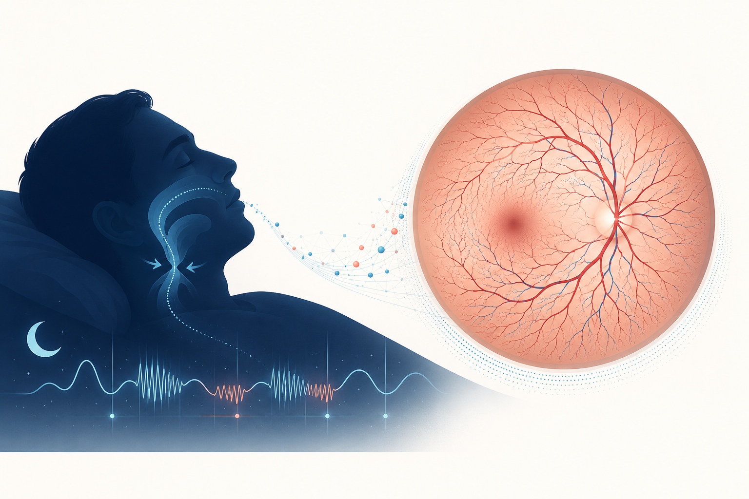



Obstructive sleep apnea (OSA) is characterized by recurrent upper-airway collapse during sleep. Although the disorder is defined by nocturnal respiratory events, its pathophysiological consequences may extend beyond the upper airway. Intermittent hypoxemia, arousal-related sympathetic activation, and hemodynamic instability have been associated with cardiometabolic, cerebrovascular, and ocular abnormalities. Whether these processes are accompanied by measurable alterations in retinal microvascular structure has not been fully established.
This study was undertaken to determine whether retinal imaging could provide a noninvasive approach to examining microvascular patterns associated with OSA severity.
The retina represents an accessible vascular bed in which microcirculatory features can be directly visualized and quantified in vivo. Previous investigations of OSA have mainly evaluated retinal vessel caliber, including arteriolar and venular diameters; however, the reported findings have been inconsistent. Because vessel caliber reflects only one component of retinal vascular morphology, retinal vascular fractal dimension (FD) was examined as a complementary measure of vascular branching complexity and density.
Linking sleep physiology with retinal imaging
The apnea–hypopnea index (AHI) is the most widely used indicator of OSA severity because it quantifies the frequency of respiratory events during sleep. Nevertheless, AHI does not fully characterize the physiological burden associated with recurrent hypoxemia or its potential effects on the systemic microvasculature.
Retinal vascular FD was therefore used to assess the overall organization of the retinal vascular network. Higher FD values generally indicate greater vascular branching complexity and density, whereas lower values indicate reduced network complexity. In contrast to isolated vessel-caliber measurements, FD provides a global description of retinal vascular architecture.
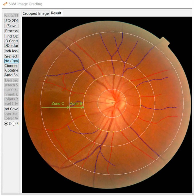
Participants who had undergone both overnight polysomnography and fundus photography during routine health examinations were retrospectively identified. Sleep recordings were evaluated by sleep specialists, and retinal photographs were analyzed using Singapore I Vessel Assessment software. This design enabled the association between AHI and retinal vascular structure to be assessed after adjustment for demographic and cardiometabolic factors. It also allowed the shape of the association to be examined and evaluated according to diabetes status.
Associations between AHI and retinal vascular structure
The analysis included 468 participants, of whom 355 met the diagnostic criteria for OSA. Retinal FD decreased across increasing AHI tertiles. After adjustment for demographic and cardiometabolic covariates, participants in the highest AHI tertile had lower overall FD and arteriolar FD than those in the lowest tertile.
This pattern warrants attention because several conventional retinal vessel-caliber measures did not show statistically significant adjusted differences across increasing OSA severity. The findings suggest that FD may provide structural information that is complementary to conventional vessel-caliber measures, rather than serving as a replacement for them.
The restricted cubic spline analyses further indicated that the association between AHI and retinal FD differed according to diabetes status. Among participants without diabetes, overall, arteriolar, and venular FD values initially declined and subsequently increased at higher AHI levels, producing a U-shaped association. In participants with diabetes, these measures declined progressively with increasing AHI. This contrast suggests that the metabolic environment may influence the retinal vascular correlates of sleep-disordered breathing.
The findings should be interpreted within the limitations of the study design. Because the investigation was retrospective and cross-sectional, temporal relationships and causality cannot be inferred. OSA frequently coexists with obesity, hypertension, diabetes, dyslipidemia, and other vascular risk factors, each of which may affect retinal vascular structure. Although relevant covariates were included in the adjusted analyses, residual confounding remains possible.
OSA beyond upper-airway obstruction
OSA is commonly approached as a disorder of recurrent upper-airway obstruction. This clinical framework remains essential, although the condition may also be associated with physiological changes involving multiple organ systems, including the microvasculature.
Retinal imaging cannot replace polysomnography and should not be considered a diagnostic test for OSA. Its value in this context lies in its potential use as a research tool for investigating vascular features associated with sleep-disordered breathing. The retina provides a noninvasive and quantifiable site at which these associations can be studied directly.
The integration of polysomnography and fundus photography also illustrates the value of linking established assessments across clinical disciplines. When sleep physiology and retinal vascular imaging are evaluated within the same analytical framework, questions concerning OSA severity, microvascular architecture, and cardiometabolic risk can be addressed more comprehensively.
Priorities for future research
Prospective longitudinal studies are required to determine whether retinal fractal measures change with variation in OSA severity, whether they are associated with subsequent cardiovascular or ophthalmic outcomes, and whether they are modified by OSA treatment. Future investigations should include more diverse populations, detailed measures of hypoxic burden and sleep architecture, and external validation across different clinical settings.
It will also be necessary to determine whether retinal vascular measures contribute information beyond established cardiometabolic risk factors. Such evidence would be needed before any role outside research settings could be considered.
Taken together, these findings indicate that the consequences of disrupted breathing during sleep may be reflected in retinal microvascular architecture. Although the retina cannot serve as a surrogate for the entire vascular system, it offers a noninvasive and measurable window through which the vascular correlates of OSA can be investigated.
This post is based on: Wang J, Chen T, Qi X, Li Y, Yang X, Meng X. Retinal vascular fractal dimension measurements in patients with obstructive sleep apnea syndrome: a retrospective case-control study. Journal of Clinical Sleep Medicine. 2023;19(3):479–490. https://doi.org/10.5664/jcsm.10370

Follow the Topic
-
Journal of Clinical Sleep Medicine
Journal of Clinical Sleep Medicine (JCSM) is the official peer-reviewed, scientific publication of the American Academy of Sleep Medicine (AASM), the leading professional society in the sleep field. The journal focuses on novel, high-impact human sleep and circadian science.