JMCR: Clinical Minute
Published in Biomedical Research
Explore the Research
XRAqyy9Z8mWa?utm_campaign=related_content&utm_source=HEALTH&utm_medium=communities
Article Highlights
- Describes a facial-nerve schwannoma presenting as an occluding external auditory-canal mass.
- Demonstrates preserved facial function despite tumor origin from the facial nerve.
- Identifies smooth posterior canal-wall erosion near the mastoid facial-nerve segment as an important CT clue.
- Shows how the lesion may mimic an aural polyp, cholesteatoma, ceruminous tumor, paraganglioma, or malignancy.
- Reinforces the risk of facial-nerve injury from an inadequately planned biopsy.
- Confirms the diagnosis histologically through Antoni A and B architecture and strong S-100 expression.
Clinical Take-Home Message
Normal facial function does not exclude a facial-nerve schwannoma.
A posterior external auditory-canal mass—particularly one accompanied by smooth canal-wall erosion—should be imaged and assessed for a possible facial-nerve origin before biopsy or excision. CT defines the bony anatomy, while MRI generally provides the best confirmation of neural origin and tumor extent.
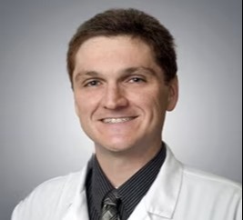
Richard Alan Rison is the Editor-in-Chief of Journal of Medical Case reports. He is also an associate neurology editor (editorial board) for BMC Neurology, and the former lead editor for case reports of BMC Research Notes (currently on the editorial board). His scholarly work focuses on medical case reporting, reporting standards, and editorial methodology. Dr. Rison participated in the development and dissemination of the CARE guidelines for clinical case reporting and has authored numerous publications addressing both neurological disorders and the role of case reports in advancing medical knowledge. Dr. Rison practices general neurology and served as the founding medical director of the PIH Health Hospital-Whittier Stroke Program and the PIH Health Hospital-Whittier Non-Invasive Vascular Laboratory, is a clinical assistant professor of neurology at the University of Southern California Keck School of Medicine and Los Angeles County Medical Center, and is a Fellow of the American Academy of Neurology, the American Neurological Association, and the American Association of Neuromuscular and Electrodiagnostic Medicine. Dr Rison is board-certified by the American Board of Psychiatry and Neurology in neurology and vascular neurology, and neurocritical care and neuroimaging by the United Council of Neurologic Subspecialties. He is also board-certified by the American Board of Electrodiagnostic Medicine in electrodiagnostic medicine. Dr. Rison is a former president of the Los Angeles Neurological Society.
Follow the Topic
-
Journal of Medical Case Reports
This journal will consider any original case report that expands the field of general medical knowledge, and original research relating to case reports.