Engineering acoustofluidic and optofluidic biosensors in medicine
Published in Bioengineering & Biotechnology, Materials, and Physics
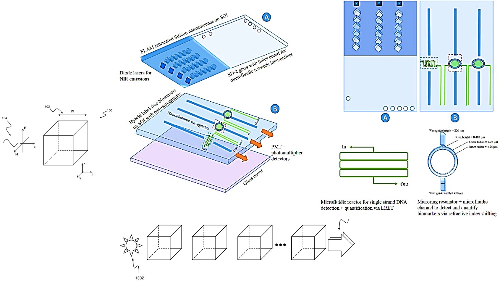
My interests on microring resonators and optofluidic devices (combining optics and microfluidics) began about a decade ago, due to their direct alignment with my research experience in physics and materials science. The advent highlighted the research possibilities of building a futuristic yet conceptual device to facilitate photonic integrated biosensors for medical diagnostics applications in biomedical engineering [Debabrata S. et al. 2015, Chandrahalim H. et al. 2015, Kues 2017]. The materials science and physics underlying the waveguide technology includes the development of novel cubic dielectric nanoparticles for photonic applications, with the capacity to precisely regulate and engineer the scattering of electromagnetic waves through sub-wavelength nanostructures for bioengineering (Figure 1) [Debabrata S. et al. 2015].
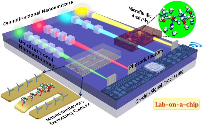
Figure 1: A schematic representation of unidirectional cubic nanoantennas inducing directionality to omnidirectional nano-emitters to precisely focus light. In concept, these ultra-nanodirectional beams can play multiple roles within, in-built lab-on-a-chip devices for microfluidic analyses, and biomolecular identification. Credit: [AAAS 2015].
Such concepts have scope in the integration of nano-lasers and photovoltaics in biomedicine [Debrata S. et al. 2013, Debabrata S. et al. 2015]. Built-in ring resonators are another inclusion to biosensor instruments that can ideally interact with another built-in waveguide and have been in development for more than two decades, while active studies of the concept only began in the lab in the early 90s, with recent improvements as inclusions within optofluidics, acoustofluidics (combining acoustics and microfluidics) and microfluidics to create advanced biosensors [Gorodetsky and Ichenko 1994]. This construct can fine-tune frequencies/resonances, to investigate exciting trajectories in the life sciences [Rabus and Sada, 2020]. Conventional ring resonators play an important role in silicon photonics, as they consist of a set of waveguides with a closed loop coupled to a light input or output. The structures support multiple resonances and in general consist of a looped optical waveguide and a coupling mechanism to access the loop, typically suited for varied functions in physics and mathematics for fibre and data center networks [Bogaerts W. et al. 2012] (Movie 1, Figure 2).

The capacity to engineer conceptual cubic dielectric nanoparticle clusters as waveguides on a chip can be brought to life via materials science, CMOS, and multiphysics for biophotonics applications (Figure 1). For instance, the aforementioned cubic nanoantennas are composed of an insulating material (rather than a conducting or semiconducting material), to efficiently direct an ultra-narrow beam of light with little to no loss via scattering or heat [Debabrata S. et al. 2015]. Such an instrument can be engineered to precisely regulate the scattering of electromagnetic waves through sub-wavelength nanostructures for precursor biological applications, to manipulate and detect bioparticles [Chandrahalim H. et al. 2015].
The conceptual micro-ring devices with cubic nano-antennas and microfluidic constructs can be engineered in-lab via materials fabrication methods such as femtosecond laser lithography-assisted micromachining (FLAM), and plasma-etching for nanofabrication (Figure 3), to combine two bioanalytical sensors within a single framework [Chandrahalim H. et al. 2015]. The space between the nanocube chain can be precisely adjusted to fine-tune the direction of the light beam. Such optical nanoantennas can have numerous applications when coupled to light-emitting structures, to form nanoscale-directed light sources for integrated optics-based biosensors, with capacity to detect proteins, DNA, antibodies, and enzymes in an advanced lab-on-a-chip system.
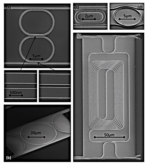
Figure 2: Examples of silicon ring resonators. (a) Double ring resonator with tuned directional coupling sections, (b) Circular ring with large coupler gaps, (c) Ultra-small racetrack ring with 1μm bend radius, (d) ring with conformal coupling sections, (e) Large folded-spiral ring. Credit: [Bogaerts W. et al. 2012].
Materials engineering acoustofluidics (combining acoustics and microfluidics) for biosensing
Multifunctional acoustic tweezers too have gained precedence as a promising technology in biosensing to form a precise, biocompatible mechanism to manipulate delicate bioparticles or biospecimen that range from the nanometer-scale to the millimeter-scale [Tian Z. et al. 2020]. The innovative acoustic tweezers use sound waves to form ‘virtual tweezers’ to precisely perform non-contact and label-free exact manipulation of bioparticles, to observe and understand cell-cell interactions, single-cell analysis, and acousto-mechanical phenotyping [Augustsson P et al. 2016, Wang H. et al. 2019]. This method can be readily adopted for cell studies, tissue regeneration and in regenerative medicine [Ozcelik A. et al. 2018].
Advances in acoustic tweezer-based strategies, however, lack reusability as they are costly and time-consuming for sterilization. This innovation also requires specialized fabrication methods for single-cell analysis experiments that rely on surface acoustic wave manipulation, to facilitate single cell-patterning experiments of ‘one cell per well’ [Collins D. et al. 2015]. Bioengineers have therefore created more recent advances to realize this concept, by leveraging acoustic tweezers for non-contact bioparticle identification within simpler frameworks such as Petri-dishes. Petri-dishes are a common and cost-effective cell culture tissue staple generally found in biomedical laboratories with scope to easily facilitate acoustic interactions. Such platforms provide a simple arrangement to generate sound waves with acoustic transducers placed outside the petri-dishes [Tian Z. et al. 2020].
Fast-forward to 2025, more than half a century after the proposal of the conceptual microring resonators in the late 1960s by Marcatili at Bell Labs[Little B et al. 2003, Marcatili E. 1969], researchers have developed a series of conceptual avant-garde instruments, including ‘acoustofluidic ring-resonators’ that are primarily based on built-in acoustic tweezing to manipulate microparticles, with significant impact to advance non-contact biosensing and mechanobiology with next-generation lab-on-a-chip techniques for cell-cell communication research (Figure 4) [Xianchen X. et al. 2024].
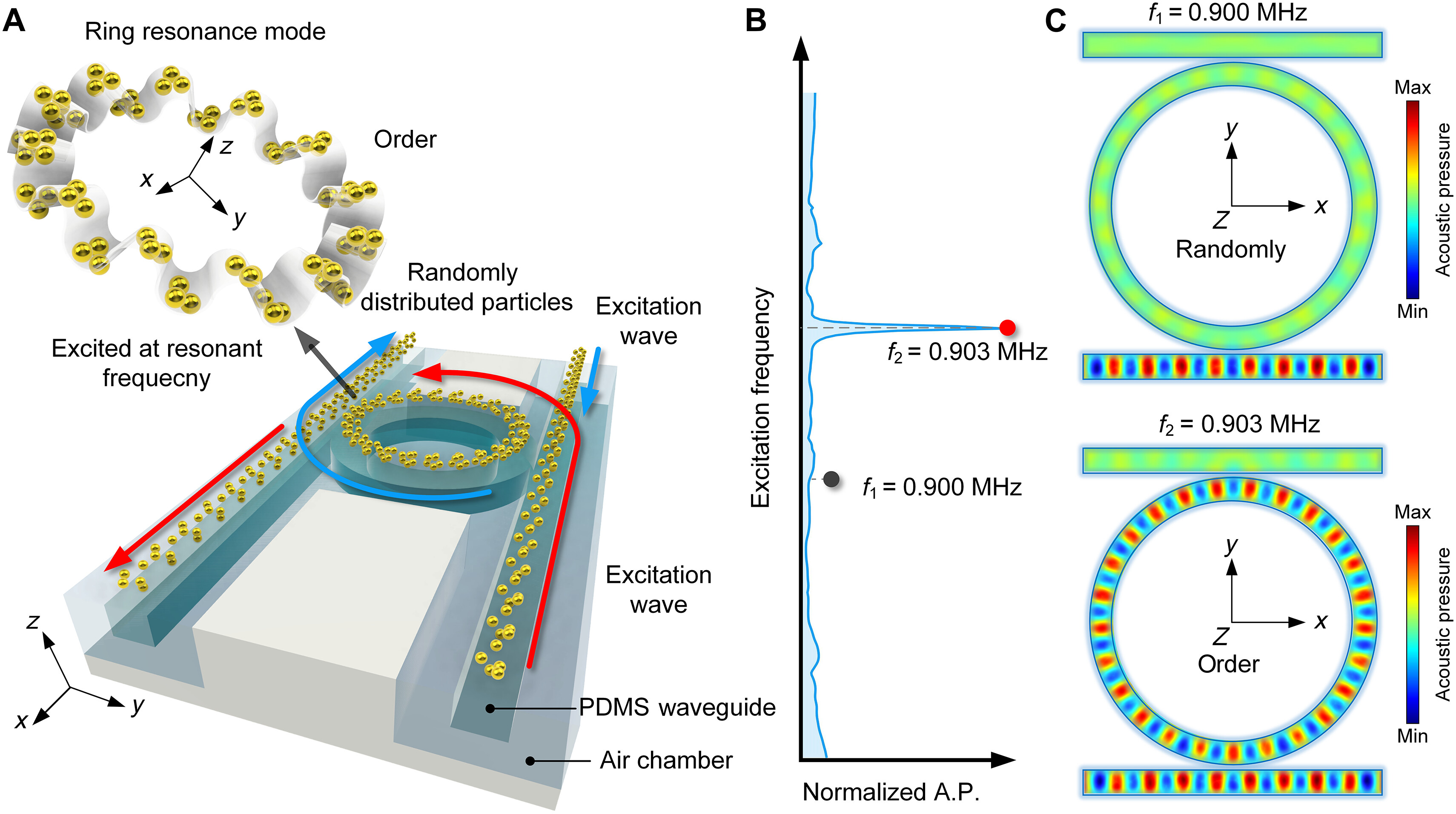
Figure 4: Acoustic tweezers via ring resonance. A) The schematic diagram shows the positioning on both sides of the Poly dimethyl siloxane (PDMS) waveguide, including clockwise and counterclockwise resonances in the ring resonator due to the red and blue input signals. B) Non-resonant and resonant frequencies of the ring resonator, and C) Acoustic field distribution at 0.9 MHz (non-resonant mode) and 0.903 MHz (resonant mode).
In concept, these prototype, and novel lab-on-a-chip microsystems can be engineered as hybrid devices with built-in analytical biosensors to function via acoustics or laser lights, while coupled to a ring resonator within a single platform for the label-free detection and quantification of oligonucleotides and genetic materials [Bhutaite U. et al. 2024]. Such devices differ from their traditional counterparts as they can, for instance, manipulate micro-sized particles through the resonance interaction of acoustic waves with a higher Q factor that far exceeds conventional acoustic transducers. Such acoustofluidic ring resonator tweezers have immense potential for advanced applications in the life sciences [Xianchen X. et al. 2024]. During its mechanism-of-action, the acoustic fields can be shaped in a frequency-dependent manner for samples to move through channels for analytical functions, such as ultrasonics integrated PCR within point-of-care devices to rapidly detect pathogens inside a single, hand-held diagnostics system [Reboud J. et al. 2012]. The ring resonator concept and its resonant frequency can be visualized with Multiphysics simulations prior to the actual experiments (Movie 2).
Bioengineering advances in acoustofluidic technology
The existing requirement for advanced bioengineering strategies in acoustic tweezer development originated to overcome limits that currently impede its workflow, which include the lack of its reusability as a single-cell pattering strategy, despite its potential for successful long-term use [Collins D. et al. 2015]. Studies have already looked at patterning human lymphocytes and red blood cells infected by the malarial parasite Plasmodium falciparum, where the analytical acoustic wavelength is in the same order as the cell dimensions [Collins D. et al. 2015]. The virtual, 3-D acoustic tweezers can also pick-up, translate and facilitate cell assemblies to create 2-D and 3-D cell assemblies in a precise, non-invasive, label-free, and contact-free method [Guo F. et al. 2016].
Recent advances in bioengineering have seen to the development of acoustic tweezers with three distinct configurations for the unique, non-contact investigation of bioparticles in a Petri-dish, as a simple and more accessible cell culture platform (Figure 5). This approach incorporates tunable standing acoustic waves, acoustic streaming vortices, and holographic interdigital transducers (IDT) for multi-configuration cell patterning, cell-cell interactions and to develop 3-D tissues [Tian Z. et al. 2020]. Bioengineers can regulate the excitation frequency, phase, and amplitude of each transducer. Other configurations of the instrument can generate an acoustic streaming vortex to drive bioparticles in a fluid layer to the vortex center and concentrate them for signal enhancement and develop large cell spheroids.
In a third configuration, by switching between different holographic interdigital transducer frequencies and designs, it is possible to achieve multiple functions such as the localized transient stimulation of cells for cell lysis, cell concentration and containment of bioparticles on a dish. The electric profile of the holographic IDT can be constructed to regulate cells and biological particles [Tian Z. et al. 2020].
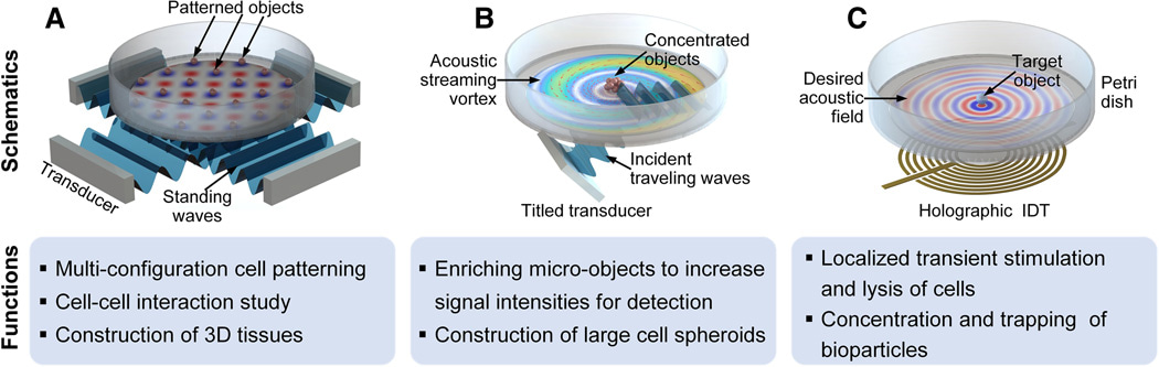
Figure 5: Using acoustic tweezer devices to regulate bioparticles in a petri-dish. A) A Petri dish is placed on top of the tweezer device compose of an array of piezoelectric transducers to generate standing acoustic waves in a fluid layer in the Petri dish. B) This device configuration presents a tilted piezoelectric transducer to generate oblique incident travelling acoustic waves. C) The third device configuration has a holographic interdigital transducer placed under the Petri dish to generate high frequency acoustic waves [Tian Z. et al. 2020].
Precise Multiphysics simulation to optimize a biology experiment
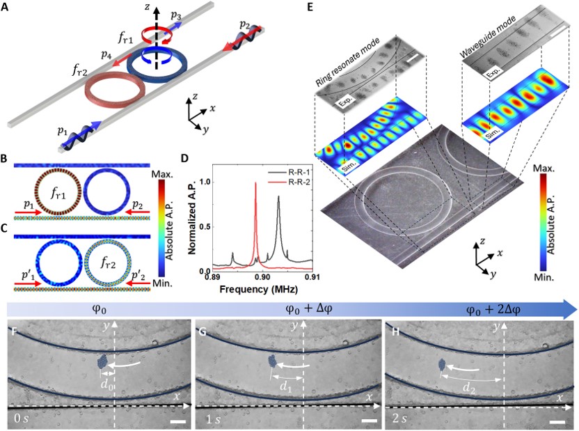
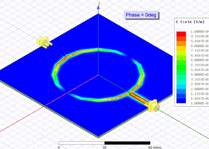
As noted earlier, even more recent advances in bioengineering have introduced acoustofluidic ring resonator tweezers with a high Q factor approximating 20 times greater than its traditional counterpart, as a conceptual, yet optimized power-efficient approach for microparticle manipulation [Xianchen X. et al. 2024]. Structurally, the ring resonator-based acoustofluidic tweezers contain two straight rectangular waveguides and a ring resonator. The waves from opposite directions propagate within the waveguide and interact in the ring resonator with a high Q factor to produce a predesigned, pure mechanical acoustic wave [Li F. et al. 2013]. With energy input, particles on the system’s surface transition from random to an ordered distribution, structured by the pressure mode to form an acoustics tweezer- particle containment effect. As with most ideas in physics, the initial concept can be simulated with COMSOL Multiphysics, based on the principles of ring resonator theory, to facilitate the ring shape and translate the concept to applications in the life sciences.
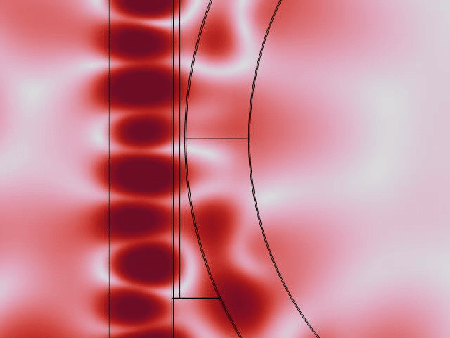
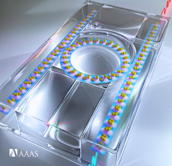
The simulation results illustrate the acoustic field distribution, to show that the acoustic field is contained in the fluid layer at the waveguide region, to facilitate bioparticle trapping inside the ring resonator (Figure 6). This simulation can be easily constructed on a polydimethyl siloxane (PDMS) surface to maintain both the waveguide and the ring resonator structure [Xianchen X. et al. 2024]. The signals within the ring resonator can be enhanced by incorporating multiple waveguides to converge on a central ring resonator (Figure 7).
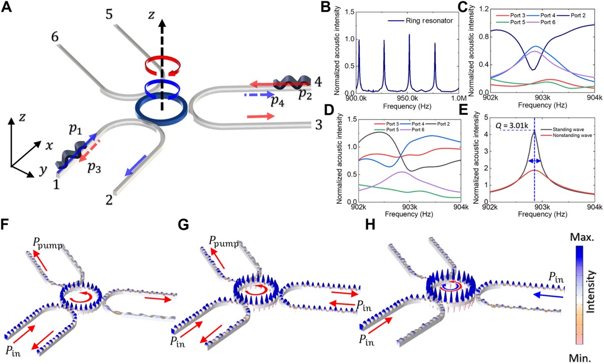
Figure 7: Three waveguides with one ring-resonator configuration for signal amplification of the ring resonator. (A-H) From the simulated experimental setup to the development of a standing acoustic wave inside the ring resonator and waveguide, with two directly opposing input signals to represent acoustic signals inside the center resonator with additional simulation results [Xianchen X. et al. 2024].
Meticulous experimental validation - regulating microbubbles in Zebrafish embryos
The ring resonator-based acoustic tweezers can be optimized with multiple PDMS rings with subtle geometric variations, to maintain distinct resonant frequencies that are suited for efficient functions. The ring resonators typically have a consistent ring diameter; and its height can be altered to adjust the resonant frequency. For instance, by selecting a specific frequency a simulation as well as an experiment can be carried out, to illustrate the capture and arrangement of PDMS particles (50 to 100 μm) within the mode shape of the ring system [Xianchen X. et al. 2024]. In this way, the acoustic waveguide and ring resonator can generate a standing wave, and researchers can manipulate particles on the ring’s surface by regulating the phase of the input signal.
The precision of the method can be illustrated with particles that move along a designated guideway within the ring-resonator. Example applications of the techniques include in vivo micromanipulation via ultrasound - demonstrated by regulating microbubbles within blood vessels for micrometer precision by using zebrafish embryos, without interfering the circulating blood vessels of the organism (Figure 8) [Jooss V. et al. 2022]. This capacity to conduct successful, high-precision in vivo acoustic regulation of microbubbles within blood vessels at micrometer precision, without interfering the circulating blood vessels can be translated to potential therapies for cancer research, neuroscience, and vascular biology [Jooss V. et al. 2022].
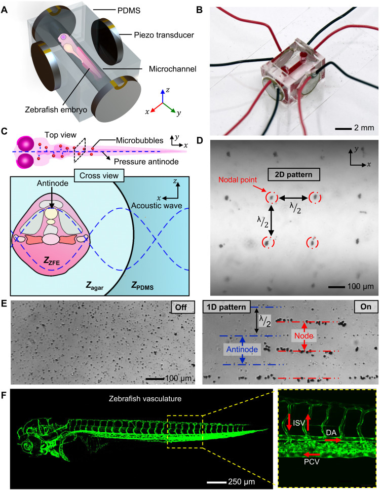
Figure 8: Experimental design and concept of the in vivo acoustic manipulation system. (A-E) Schematic of the in vivo acoustofluidic device, a micrograph of the in vivo acoustic manipulation chamber, and a schematic of the acoustic wave to visualize a zebrafish embryo. (D-F) The 2-D lattice-like acoustic pattern is produced inside the microchannel by the acoustic system, fluorescence image of a zebrafish embryo expressing green fluorescent protein (GFP) in endothelial cells highlight its vasculature network to indicate the direction of blood flow [Jooss V. et al. 2022].
Optimizing the ring-resonator and acoustofluidic tweezer combination
The capacity for a sound wave to travel around the ring resonator is similar to an already existing concept known as whispering gallery waves, first discovered by Lord Rayleigh in 1878, to describe the curvilinear propagation of sound waves around a cathedral dome [Kfir O. et al. 2020, Yu et al. 2021]. This concept is also applicable to light waves as well, when they are continuously reflected along the closed concave surface of an optical cavity built with a material with high refractive indices, such as a silicon or a glass microsphere in optofluidics. A standing wave can be developed in the waveguide to facilitate the containment of microparticles within the device, this can be accomplished when acoustic tweezers are combined with ring-resonators within the same instrument, and an incoming wave happens to be outside the resonant frequency. As the frequency approaches resonance, this gives rise to a strong standing resonance for particle containment within the ring. The designed combination of the ring-resonator and acoustic tweezers have impact across biosensing and drug delivery applications. Existing strategies have already detailed the label-free detection of ovarian cancer biomarkers by using whispering gallery mode imaging methods [Huckabay et al. 2013]. Furthermore, asymmetrical microring resonators based on whispering gallery modes can detect glucose concentrations as biosensors in the field of medical chemistry [Zhang P. et al. 2018]
Movie 3: Representing the acoustic pressure distribution on the ring and waveguide. As the frequency approaches resonance, the acoustic pressure on the ring intensified, reaching its peak at the resonant frequency. The waveguide facilitates the transmission of acoustic waves, while the ring experiences enhanced pressure due to the resonance to showcase the dynamic behavior of the system at this critical frequency [Xianchen X. et al. 2024].
The biocompatible, non-contact techniques have further practical applications for in vivo investigations, as well as quantum sensing, medical diagnostics, bioanalytical chemistry, and in materials science [Wang C. et al. 2021]. Most acoustic tweezers require a strong input power to generate a sufficient force, which can result in unwanted heat generation and other technical difficulties that necessitate high-performance transducers. This technicality can be negated by introducing high Q resonances to achieve strong wave-particle interactions, with a moderate input of power.
In the realm of built-in acoustofluidic tweezers therefore, a high Q factor signifies superior performance where the field intensity depends on pairs of input signals. To optimize the Q factor, multiple waveguides can converge to a central ring resonator to heighten the quality of the signal and advance the capacity of the acoustic tweezers for applications in precision cell sorting, and as drug delivery platforms (Figure 7, movie 3). The acoustic pressure distribution on the ring and waveguide is better visualized in movie 3.
Developing a conceptual device based on the synergy between physics and bioengineering
In this way, ring resonance physics and bioengineering can be integrated for particle manipulation to create novel acoustofluidic ring-resonator based tweezers with a high Q factor (Figure 9). These concepts can yield promising outcomes to overcome existing limits of optical tweezers to precisely regulate micron-sized particles, sort out bacterial viability on a Petri-dish, or on a microfluidic chip, based on in lab acoustofluidics [Ericsson M. et al., 2000]. Cutting-edge methods have shown how bioengineers and physicists have meticulously validated the combined effects of ring resonators and acoustic tweezers, to demonstrate their effectiveness in the lab. To establish the concept, it is necessary to develop a standing wave pattern along the resonant ring path and incorporate acoustic lenses to create pressure antinodes for particle mixing.
Example studies imply a transformative trajectory for acoustic tweezers by positioning them as versatile tools to precisely regulate the particles. The high Q resonances can facilitate strong wave-particle interactions, with a moderate power input to ensure the field is strong, with minimal issues of heat generation, and bubble formation. High sensitivity biosensing applications of the instrument can be coupled with single-cell level interactions for profound technical impact of acoustofluidics with biological samples in the life sciences. The pressure levels within the ring resonator approximate 1 MPa; a value that is typically below the known threshold to cause cell damage (Movie 2).
In addition to the features detailed herein, the novel device can be designed to be frequency selective and function at a target frequency with lower voltage input, for safe use of the instrument with living cells. The innovative platform can facilitate contact-free biological particle manipulation on an organ-on-a-chip instrument, with fine-tuned acoustic pressure levels, to retain the viability of living cells while serving as a foundation for further advancements. The capacity for the robust realization and implementation of these inventions in physics within the life sciences via materials engineering methods will have tremendous impact in the dynamic field of biomedical research.
Header Image: An example of a chip-scale laser created inside an optical cavity to emit light with a fundamental linewidth at a frequency suited for biosensing and transdisciplinary scientific applications. Image credit: [Gundavarapu S. et al. 2018].
References
- Debabrata S. et al. Optically resonant magneto-electric cubic nanoantennas for ultra-directional light scattering, Journal of Applied Physics, 2015.
- Chandrahalim H. et al., Monolithic Optofluidic Ring Resonator Lasers Created by Femtosecond Laser Nanofabrication., Lab-on-a-Chip, 2015
- Kues M. et al. On-chip generation of high-dimensional entangled quantum states and their coherent control, Nature, 2017
- Debrata S. et al. Optimized gold nanoshell ensembles for biomedical applications, Springer Nature Link, 2013
- Gorodetsky and Ichenko, High-Q optical whispering-gallery microresonators: precession approach for spherical mode analysis and emission patterns with prism couplers, Optics Communications, 1994
- Rabus and Sada, Integrated ring resonators, Springer Nature Link, 2020
- Bogaerts W. et al. Silicon Microring Resonators, Laser and Photonics Reviews, 2012
- Tian Z. et al. Generating multifunctional acoustic tweezers in Petri dishes for contactless, precise manipulation of bioparticles, Science Advances, 2020
- Augustsson P et al. Iso-acoustic focusing of cells for size-insensitive acousto-mechanical phenotyping, Nature Communications, 2016
- Wang H. et al. A continuous-flow acoustofluidic cytometer for single-cell mechanotyping, 2019
- Ozcelik A. et al. Acoustic tweezers for the life sciences, Nature Methods, 2018
- Collins D. et al. Two-dimensional single-cell patterning with one cell per well driven by surface acoustic waves, Nature Communications, 2015
- Little B., Advances in Microring Resonators, Optical Publishing, 2003
- Marcatili E. Bends in optical dielectric guides, Nokia Bell Labs, 1969
- Bhutaite U. et al. Photon-efficient optical tweezers via wavefront shaping, Science Advances, 2024
- Xianchen X. et al. Acoustofluidic tweezers via ring resonance, Science Advances, 2024
- Reboud J. et al. Shaping acoustic fields as a toolset for microfluidic manipulations in diagnostic technologies, Proceedings of the National Academy of Sciences of the United States of America, 2012
- Guo F. et al. Three-dimensional manipulation of single cells using surface acoustic waves, Proceedings of the National Academy of Sciences of the United States of America, 2016
- Li F. et al. Acoustic whispering gallery mode coupling with Lamb waves in liquid, Sensors, and Actuators A: Physical, 2013
- Jooss V. et al. In vivo acoustic manipulation of microparticles in zebrafish embryos, Science Advances, 2022
- Kfir O. et al. Controlling free electrons with optical whispering-gallery modes, Nature, 2020
- Yu et al. Whispering-gallery-mode sensors for biological and physical sensing, Springer Nature Link, 2021
- Huckabay H. et al. Label-free detection of ovarian cancer biomarkers using whispering gallery mode imaging, Biosensors and Bioelectronics, 2013
- Zhang P. et al. Asymmetrical microring resonator based on whispering gallery modes for the detection of glucose concentration, Optik, 2018
- Wang C, et al. Continuous monitoring of deep-tissue hemodynamics with stretchable ultrasonic phased arrays, Nature Biomedical Engineering, 2021
- Ericsson M. et al., Sorting out bacterial viability with optical tweezers, Journal of Bacteriology, 2000
- Gundavarapu S. et al. Sub-hertz fundamental linewidth photonic integrated Brillouin laser, Nature Photonics, 2018
I am an interdisciplinary researcher with a strong commitment to bioengineering, biochemistry, organ-chips and molecular biology. As well as biomechanics and biomineralization in the broader context of medicine. I completed my PhD at the University of Sydney Australia in December 2016, and travel often, find me on Twitter and irl.


Please sign in or register for FREE
If you are a registered user on Research Communities by Springer Nature, please sign in