Evaluating the Impact of Contrast Agents on the Mechanics of the Tendon-Bone Interface
Published in Bioengineering & Biotechnology, Materials, and Anatomy & Physiology
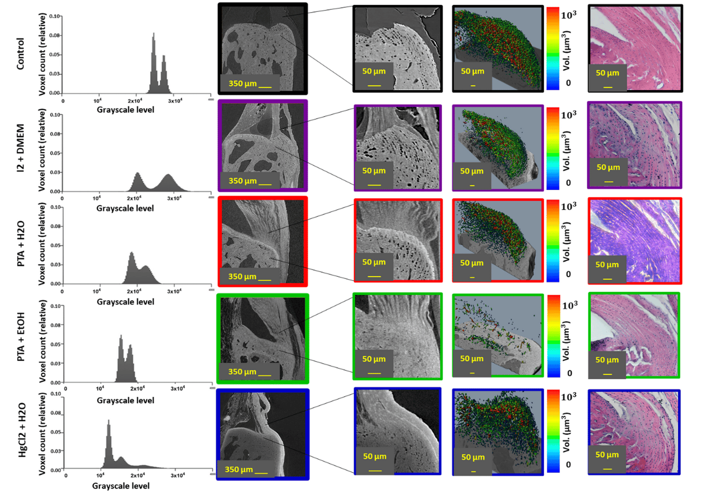
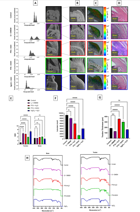
We often use contrast-enhanced micro-CT imaging to peer inside biological tissues like the tendon-to-bone interface, leveraging agents such as iodine, PTA, or mercury chloride to improve visibility. But we realized that we knew little about how these contrast agents might be altering the tissue itself, aspects like thickness, mechanical properties, and critical microstructures were overlooked.
Could these agents be reshaping the very structures we hoped to observe? We aimed to systematically assess how four commonly-used staining solutions (Iodine in DMEM, PTA in water, PTA in ethanol, and HgCl₂ in water) affect not only image quality but also the micro- and nano-mechanical integrity of the murine Achilles tendon-to-calcaneus interface. We combined high-resolution in situ micro-CT, digital volume correlation, nanoindentation, and structural analysis to gain a comprehensive view
Key Findings: All contrast agents significantly altered mechanical behaviour and structural features of the enthesis. No contrast agent perfectly preserved both tissue structure and mechanical integrity. Among the four Iodine in DMEM offered the best compromise between clear visualization and preservation of mechanical properties.
"Why it matters"
With structural-function relationships at the heart of biomechanics, understanding how imaging preparations skew observations is vital. Researchers relying on contrast-enhanced imaging for strain analysis, tissue modelling, or mechanobiology must know the trade-offs. Our work makes clear which agents preserve fidelity, and which introduce bias.
The research underscores the need for methodological rigor and critical evaluation of sample preparation protocols in biomechanical imaging. We hope this study inspires the development of improved staining protocols and agents that enhance visibility while minimally altering tissue microarchitecture.
Follow the Topic
-
Scientific Reports
An open access journal publishing original research from across all areas of the natural sciences, psychology, medicine and engineering.

Related Collections
With Collections, you can get published faster and increase your visibility.
Infectious disease diagnostics
Publishing Model: Open Access
Deadline: Sep 23, 2026
AI in Education
Publishing Model: Open Access
Deadline: Oct 09, 2026


Please sign in or register for FREE
If you are a registered user on Research Communities by Springer Nature, please sign in