Highlighting three posters from the 15th Workshop of the European Network of Breast Development and Cancer: Methods in Mammary Gland Biology and Breast Cancer Research
Published in Cancer and Cell & Molecular Biology

We are thrilled to congratulate the winners of the four Best Poster prizes, each named in honor of legendary figures in the field. Award winners:
- The Glukhova-Medina Prize: Marika Caruso received this award for her poster titled "Dynamic organoid models of mammary gland remodeling to study the effect of the estrus cycle, pregnancy, and involution on pre-oncogenic cells."
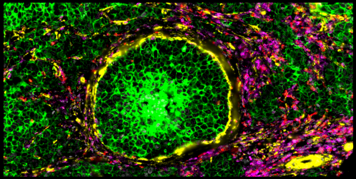
- The Hynes-Streuli Prize: Helga Bergholtz won for her work on "Single-cell spatial proteomics of HER2-positive ductal carcinoma in situ."
- The Werb-Cardiff Prize: Runyu Xia was recognized for her poster "Mouse mammary gland atlas reveals effects of age, parity, and BRCA1 loss on cell composition and immune checkpoints."
- The Watson-Rosen Prize: Hugo Croizer's research on "Deciphering the spatial landscape and plasticity of immunosuppressive fibroblasts in breast cancer" earned him this accolade.
As part of their Best Poster Prize from the Journal of Mammary Gland Biology and Neoplasia, we are pleased to present a brief overview of three of these emerging researchers and their groundbreaking work in their own words.
Marika Caruso
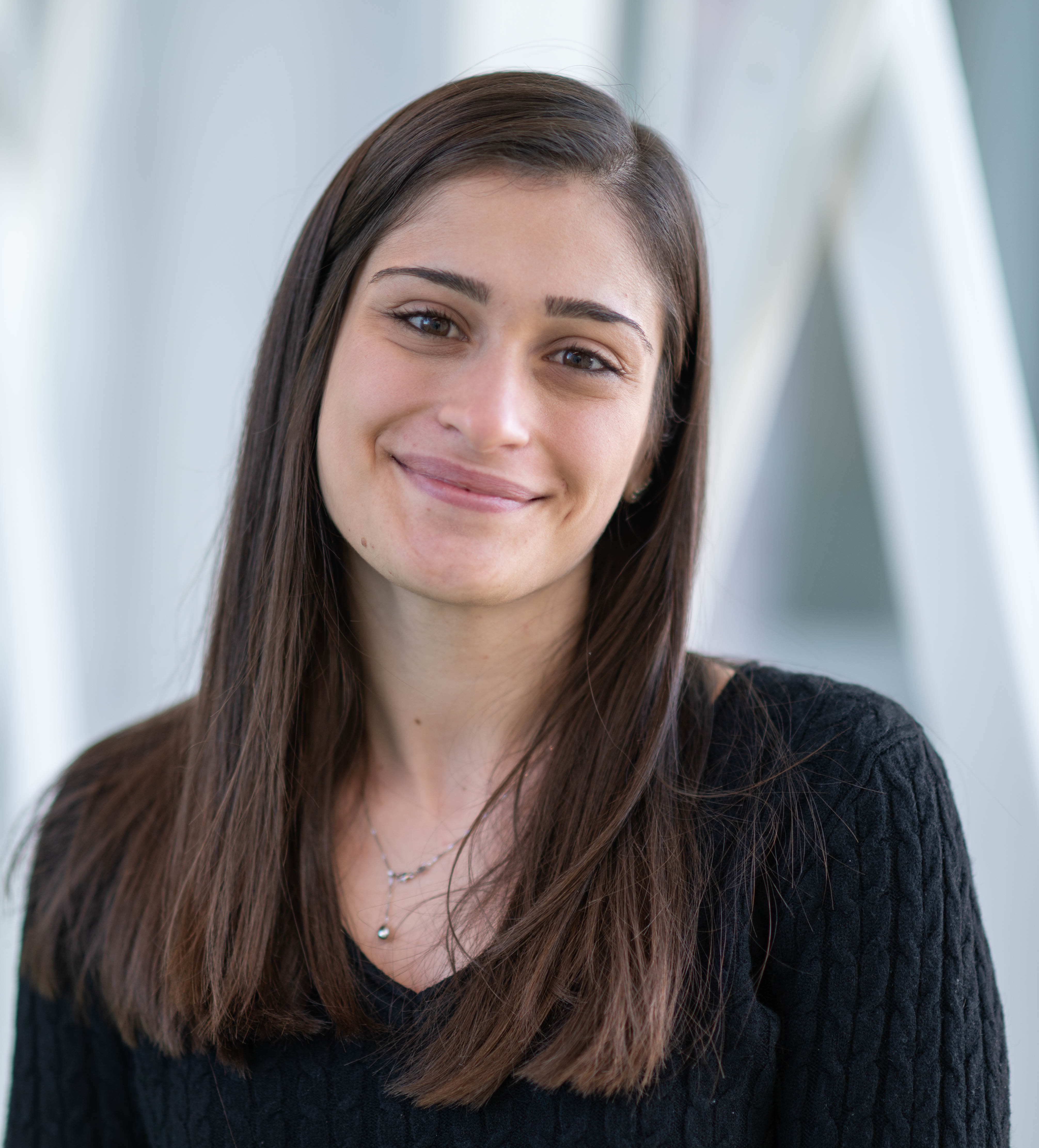
PhD Student, VIB-KU Leuven Centre for Cancer Biology, Leuven, Belgium
The mammary gland is a highly dynamic organ and it continuously remodels at each estrus cycle, during pregnancy, lactation and involution. The Scheele lab aims to reveal how this physiological tissue remodeling of the mammary gland can impact on pre-oncogenic cell behavior and how different remodeling stages can promote or protect during the different steps of tumorigenesis.
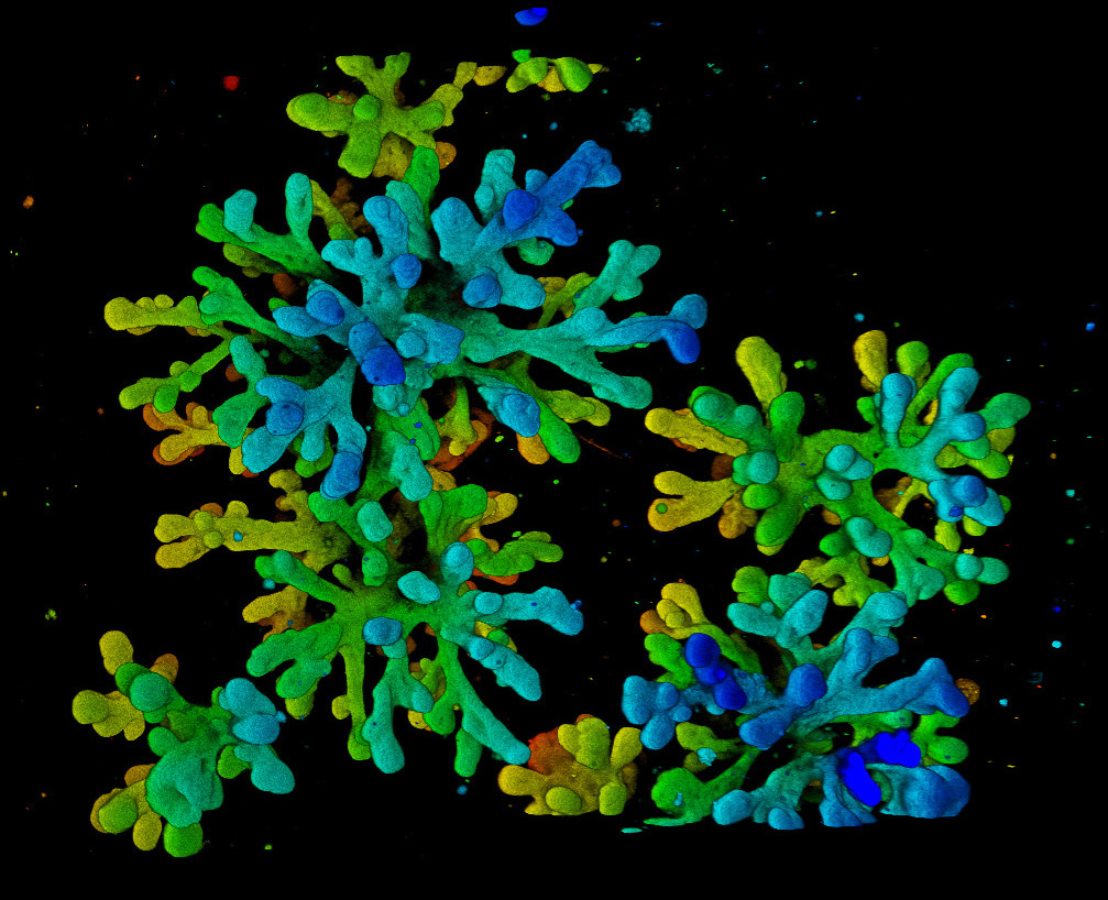
To this aim, we developed a dynamic mouse mammary gland organoid model that forms a complex network of branches and mimics the main morphological and molecular changes occurring during estrus cycle, pregnancy, lactation and involution. We are now using this dynamic organoid model to study the effect of pregnancy and involution remodeling on pre-oncogenic cells bearing mutations commonly found in breast cancer.
Runyu Xia
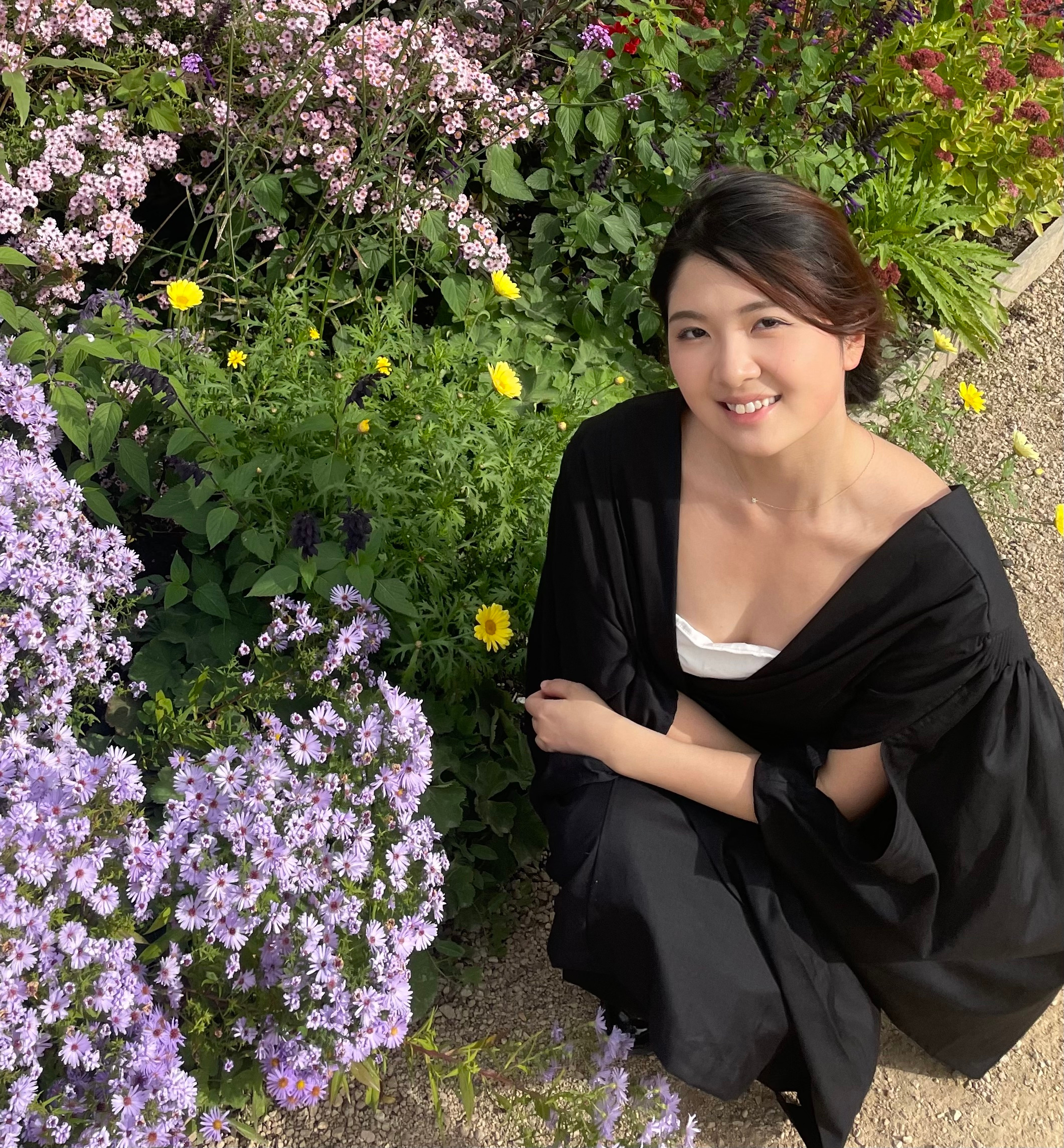
PhD student, University of Cambridge, Department of Pharmacology, Cambridge, United Kingdom
Our laboratory, led by Prof. Walid Khaled, is dedicated to uncovering the early cellular and molecular events that drive tumor initiation and development. Specifically, we investigate how the cell of origin influences the differentiation pathways of emerging tumor cells and alters the microenvironment, thereby promoting tumor growth and immune evasion. By identifying these cellular and molecular changes, we aim to develop effective early detection and intervention strategies.
The mammary gland undergoes significant transformations during various life stages, influenced by multiple risk factors. The intricate interplay between these risk factors and the diverse cell types within the mammary gland contributes to breast cancer development, though the precise nature of these interactions remains poorly understood.
To address this challenge, we have created a Mouse Mammary Gland Atlas by integrating four of our single-cell RNA sequencing (scRNA-seq) datasets. This atlas encompasses 73 samples representing a broad spectrum of risk factors, including age, parity, and oncogenic mutations such as BRCA1 knockout. Our cell type composition analysis revealed distinct changes in epithelial, fibroblast, and immune cell populations associated with age, parity, and BRCA1 knockout. Additionally, our analysis of immune checkpoints indicated age and BRCA1 LOF associated upregulation of exhaustion markers in CD8 T cells.
We hope that the findings from the mouse mammary gland atlas will provide insights into the complex interplay between aging, parity, and BRCA1 loss-of-function in shaping the mammary gland microenvironment, advahttps://ous-research.no/sorlie/ncing our understanding of breast cancer development.
Helga Bergholtz
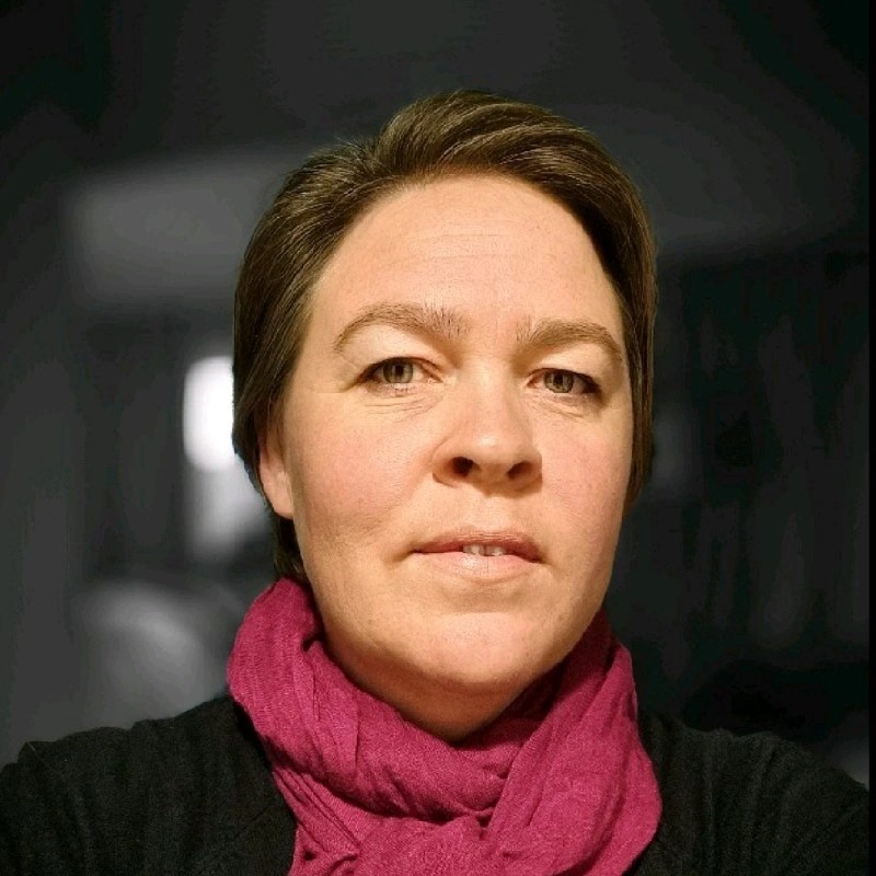
Researcher, Breast tumor evolution group, Institute for Cancer Research, Oslo University Hospital, Norway
In the Breast tumor evolution group, led by Prof. Therese Sørlie, we aim to understand breast tumor initiation and progression. We have a special interest in understanding the transition from in situ (intraductal) breast carcinomas to invasive carcinomas. We use material from patient cohorts and animal models and apply high-throughput genomic technologies, functional assays, lineage-tracing, in situ hybridization techniques and statistical and bioinformatics methods.
Recently, we have applied spatial transcriptomics and single cell spatial proteomics on intraductal and invasive carcinomas from patients with the aim of understanding the mechanisms of how breast tumors escape the milk ducts and become invasive. Spatial omics technologies enable us to obtain information from both the cancer cell compartments and the microenvironment down to single cell level.
In our study, we found distinct differences in the immune cell composition both between tumors of different molecular subtypes and between intraductal and invasive tumors. In tumors of the HER2-positive subtype, B cells and CD4+ T cells were more abundant in the intraductal tumors, while CD8+ T cells were more frequent in invasive tumors. Interestingly, tertiary lymphoid structures (lymphoid tissue embedded within the tumor tissue) were more abundant in intraductal tumors. This study points to the microenvironment as a potential regulator of invasion and that immune cells, and B cells in particular, may play central roles in breast tumor progression.



Please sign in or register for FREE
If you are a registered user on Research Communities by Springer Nature, please sign in