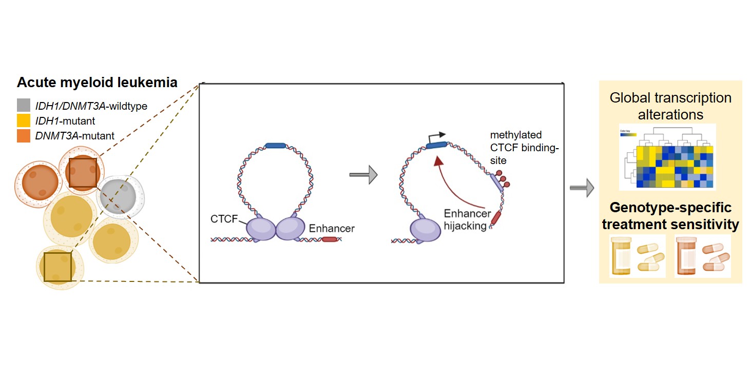

When we think about DNA, we often picture the familiar double helix - a long sequence of genetic information that contains the instructions for our cells. But inside the nucleus, DNA is anything but linear. It is folded, packaged and organized into a complex three-dimensional structure. This organization brings distant regions of the genome into close contact and can determine which genes are active and which remain silent. So, could this three-dimensional organization also help explain why different forms of acute myeloid leukemia (AML) behave differently? That was the question that motivated our study, “Alterations in 3D-DNA architecture drive unique leukemic profiles in IDH1- and DNMT3A-mutant acute myeloid leukemia.”
AML is a genetically diverse disease. Among the mutations frequently found in AML are alterations in the epigenetic regulators IDH1 and DNMT3A. Both mutations can change DNA methylation and gene regulation, but they are associated with distinct biological and clinical features. We therefore wondered: if these mutations can change the epigenetic landscape, could they also change the three-dimensional architecture of the genome? And could these structural changes contribute to the specific characteristics of the leukemia cells?
To address these questions, we combined two approaches that provide complementary information. Firstly, we used Hi-C, a chromosome-conformation-capture technique that allows us to study interactions between different regions of the genome on a genome-wide scale. But how can DNA regions that are far apart in the linear genome interact with each other? The answer is a mechanism that involves CTCF, a protein that binds to specific CTCF anchor sites on the DNA. When CTCF binds at there, it can bring two sites closely together, forming a DNA loop containing all the DNA in between. In this way, regulatory elements and genes that are normally far apart on the DNA sequence can physically interact and that interactions can regulate gene expression. Second, we used RNA sequencing to determine whether changes in this 3D architecture also translate to a changed gene expression landscape in AML with IDH1- or DNMT3A mutation. For both, we used engineered cellular models carrying either the IDH1 p.R132H or DNMT3A p.R882H mutation and also compared our findings with primary AML samples to see whether the patterns observed in the model systems could also be found in patient-derived leukemia cells.
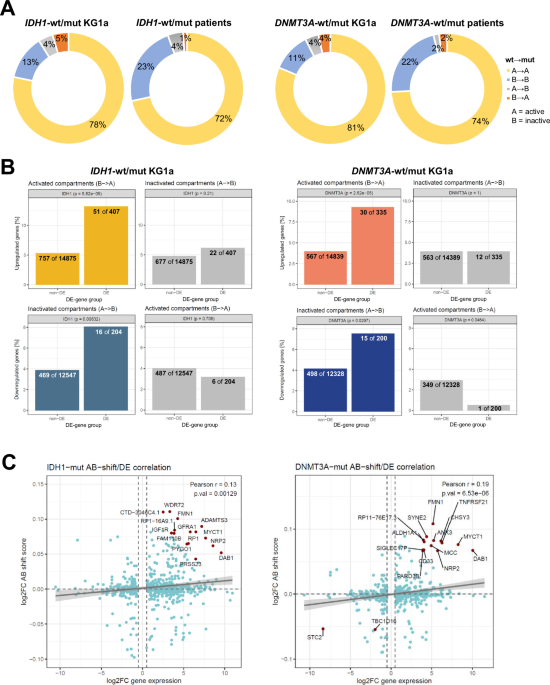
Our analyses showed that both IDH1 and DNMT3A mutations were associated with changes in 3D genome organization. Some of these alterations were shared between the two AML subtypes, others were clearly different. We observed large-scale changes affecting whole chromatin compartments as well as changes in more specific DNA-loop interactions. Importantly, these structural changes were indeed associated with differences in gene expression. In other words, the presence of either mutation specifically influences the way the genome is organized in the nucleus and that globally affects gene expression patterns.
One of the most interesting examples emerged in IDH1-mutant AML. We identified altered DNA looping around the IGF1R gene. The resulting difference in interactions with other regulatory elements was associated with increased IGF1R expression, suggesting that the altered 3D architecture was contributing to the signaling characteristics of IDH1-mutant leukemia cells. But does this also have a functional consequence? To test that, we treated the cells with the IGF1R inhibitor BMS-754807 and observed that IDH1-mutant AML cells did show a specific sensitivity to this drug. Interestingly, the benefit of BMS-754807 was also clearly visible when combined with the IDH1 inhibitor ivosidenib. For us, this was an important finding: a change in genome architecture could be connected to gene regulation and, ultimately, to a potential therapeutic vulnerability.
The DNMT3A-mutant cells on the other hand told a different story. Though there were architectural changes shared with IDH1-mutant AML, the two types of mutations also produced distinct patterns of genome organization and gene expression. In the case of mutated DNMT3A, these changes reflected in an increased sensitivity to the p38/MAPK inhibitor ralimetinib - also a potentially new therapeutic option.
These findings suggest that both IDH1 and DNMT3A mutations have distinct genomic landscapes, and that these landscapes may contribute to distinct signaling dependencies. This adds another layer to how we understand AML. But even more importantly, unraveling this new regulatory layer helped us discover new treatment options for both AML entities that have not been described before. So in the end, the key to understanding leukemia and discovering new therapy targets may sometimes be found by staying in the loop.

Since now 7 years, I work as research scientist at University Hospital Schleswig-Holstein in Kiel, Northern Germany. My research focuses on leukemia - on mechanisms that drive its development, why it comes back, and why some therapies work for some patients but not others. In my eyes, research does not end with a publication though but new findings need to be communicated to the public - the people it affects, the people whose tax money funds it and the people that may become the next generation of scientists. Both go closely together and both are what motivates me in my work as scientist.