Animating Multi-Organ Engineering Protocols
Published in Protocols & Methods

Takebe Lab investigates early organogenesis and pathogenesis of the digestive tract by leveraging human stem cell-derived miniature organ system, aka organoids. We recently established protocols for the generation of hepato-biliary-pancreatic organoid (HBPO) from human pluripotent stem cells, originally published in 2019 (1) with a follow up paper now published at Nature Protocols (2).
We herein discuss our communication design strategies with two inter-related but different articles. Given the evolving journal lineups and preprint servers, it is becoming more critical to convey ideas at glance to attract attention and improve readability at glance. To this end, we recently established a communication design center in the Yokohama City University (YCU-CDC), wherein graphic/web designers and copywriters are joining as a staff member.
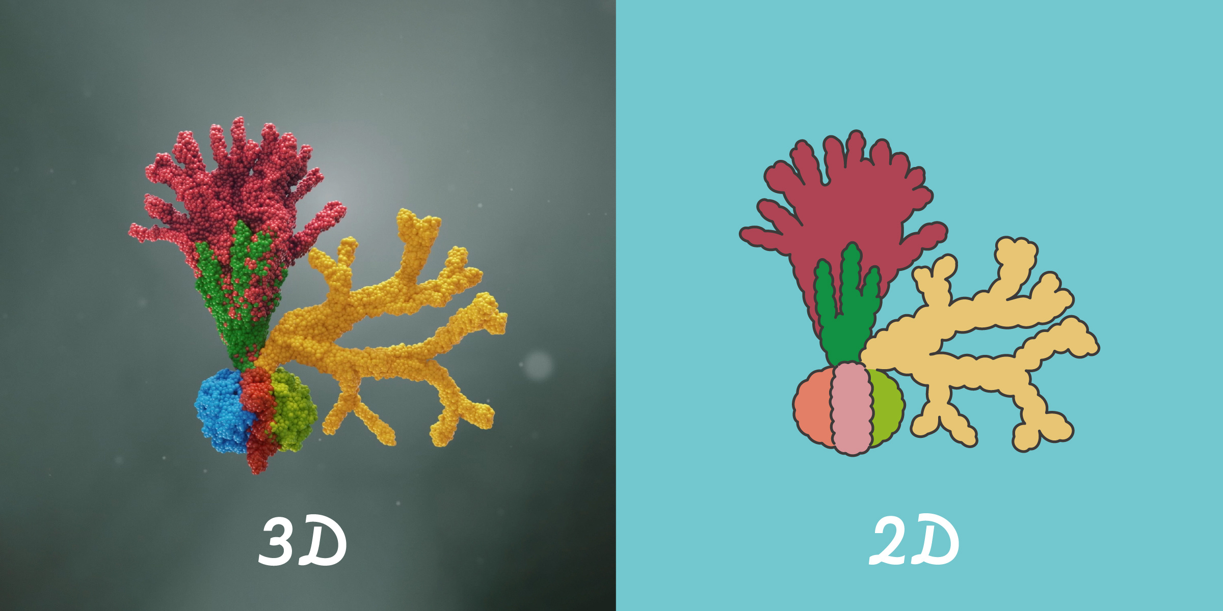
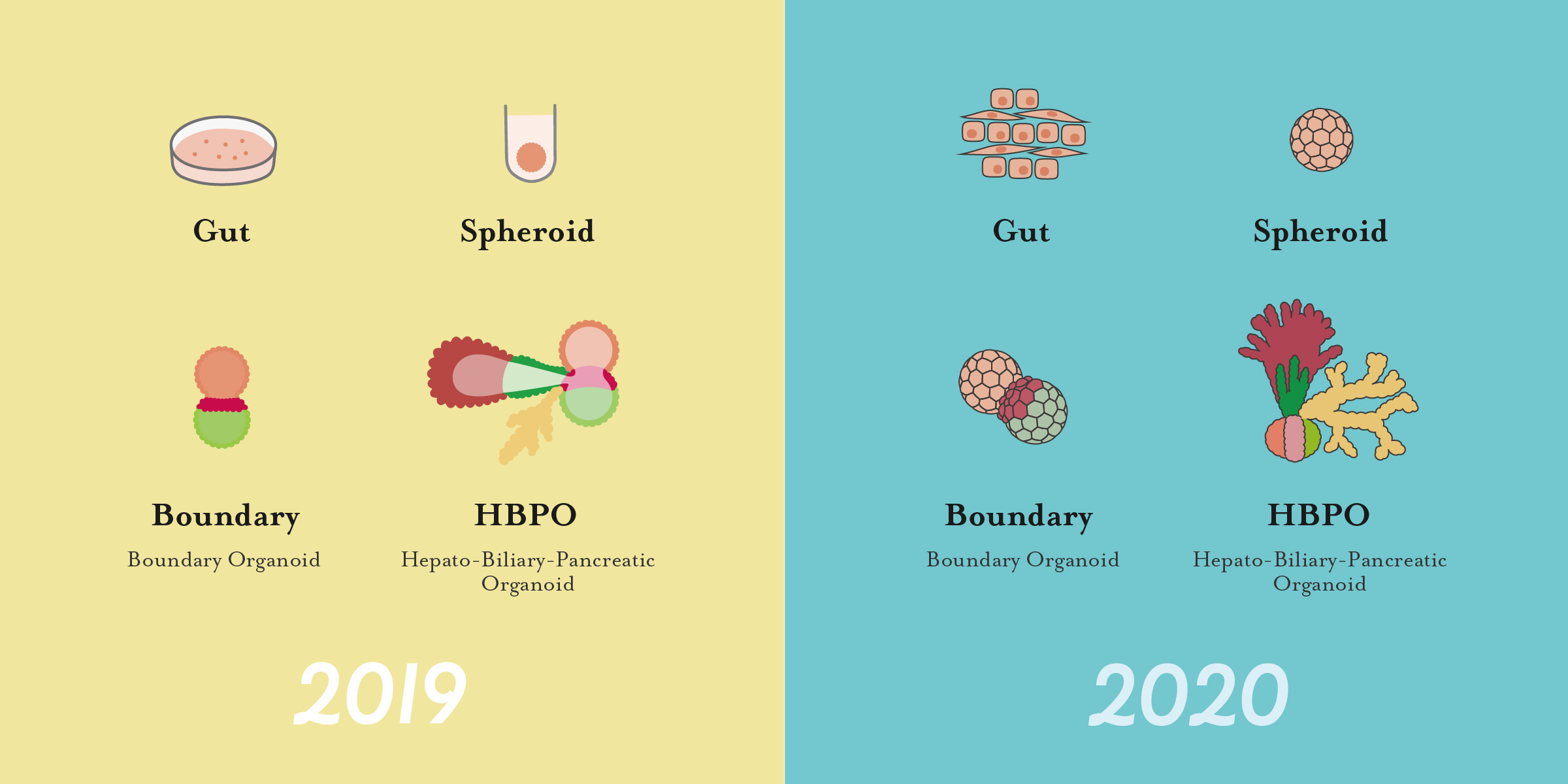
Key ideas in Nature 2019 (CG) vs Nature Protocols 2020 (Graphic) paper
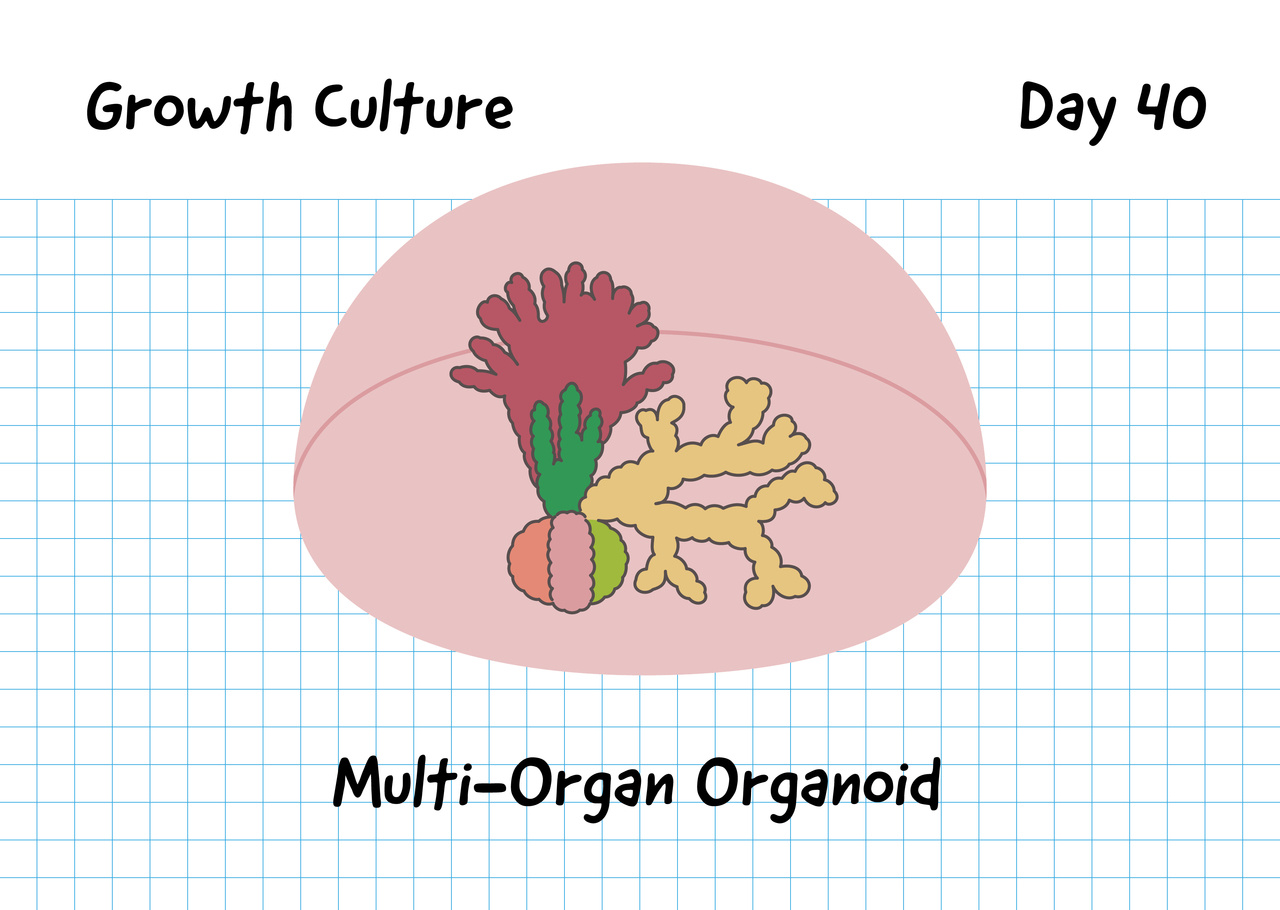
In the original research article, the main purpose of visual items is to represent conceptual advance and make a strong impression: multi-organ engineering in the organoid system. In contrast, the follow up protocol paper in 2020 was intended to convey the granular details of the protocols with some technical tips at glance. Therefore, we applied different communication design strategies in creating graphic and video materials to facilitate the readability and visibility.
The premise of a protocol paper is to make sure the procedures to be followed by students, staff scientists and technicians to obtain the results of the research published in the paper. The experimental technician is also the recipient of the information. That being said, we decided to base on illustration based materials with the intention of simplifying the experimental process.

Realistic vs Simplistic
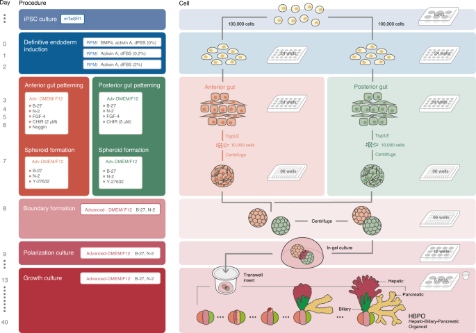
For example, our multi-organ engineering protocols are initiated by the "patterning" event upon boundary culture between two stem cell-derived spheroids with distinct regional identity: anterior and posterior gut endoderm spheroids. The surface of the anterior/posterior gut spheroids was illustrated to be more realistic by denoting the shape and ratio of each cellular component. As the well-plate format and some of the experimental instruments are important, we noted this in Figure1. Equally important is the composition of the medium and each key components were overlaid on the illustration.

Emulating the "calendar visuals"
Generally stem cell differentiation protocols require very strict time-dependent operation. Hence, we adopted a "calendar-like table" motif that was originally used in Ref3 to organize information to support the reader's interpretability. For example, at day 13 column; Boundary organoids embedded in Matrigel are transferred to trans-wells and incubated in 6-well plates with Advanced-DMEM/F12 medium until day 40 (see Ref2). The readers will be able to easily understand this sequence of events with the calendar motif. The "color bar" helps the reader to match up the day-to-day medium formulation as well as experimental manipulation.
The animated abstract
We also implemented the animated abstract as part of supplementary information, as the amount of information that is thrown from the static figures is very limited. While figures can show the snapshots of each operation in 30,000 foot view, the animation can highlight the progressive and gradual changes of each element: cellular morphology, the precise plate selection (e.g. U-bottom or Flat-bottom) and the instruments used. For example, careful attention needs to be paid on the pipette tip selection for spheroid transfer process, which is the wide bore tip. This can be easily illuminated in the video. Similarly animation can show the cellular shape conversion from a circle to a square in earlier differentiation process, and the time-dependent boundary organoid growth in later stages, which are not possible to visualize in a still image.
Although scientific writing and dataset still stand as a most important element, we think that the disseminating ideas requires design of communication. We'd like to acknowledge Asuka Kodaka for all design ideas and illustration and the other members at Yokohama City University Communication Design Center (YCU-CDC).
References
1. Modelling human hepato-biliary-pancreatic organogenesis from the foregut–midgut boundary
H Koike, K Iwasawa, R Ouchi, M Maezawa, K Giesbrecht, N Saiki, ...Nature 574 (7776), 112-116.
2. Engineering human hepato-biliary-pancreatic organoids from pluripotent stem cells
Hiroyuki Koike, Kentaro Iwasawa, Rie Ouchi, Mari Maezawa, Masaki Kimura, Asuka Kodaka, Shozo Nishii, Wendy L. Thompson & Takanori Takebe
Nature Protocols (2021)
3. Generation of multi-cellular human liver organoids from pluripotent stem cells
Wendy L Thompson, Takanori Takebe. Methods Cell Biol (2020);159:47-68. doi: 10.1016/bs.mcb.2020.03.009.
Follow the Topic
-
Nature Protocols

This journal publishes secondary research articles and covers new techniques and technologies, as well as established methods, used in all fields of the biological, chemical and clinical sciences.


Please sign in or register for FREE
If you are a registered user on Research Communities by Springer Nature, please sign in