Bottom-up engineering a pathological cell signaling pathway on an organ-on-a-chip instrument
Published in Bioengineering & Biotechnology

Following up from my previous article on organ-on-a-chip technology published here on Nature Bioengineering, in December 2016. I am gladly revisiting the concept since it intersects with my ongoing research focus, again at the intersection of medicine and bioengineering. While I am still as fascinated with the experimental detail (Bhatia 2014) surrounding the development of microfluidic cell culture devices in my research career, as I was at the completion of my PhD (in September 2015, followed by graduation in December 2016). The concept of organ-on-a-chip itself has come a long way, to ask and answer key questions in the life sciences.
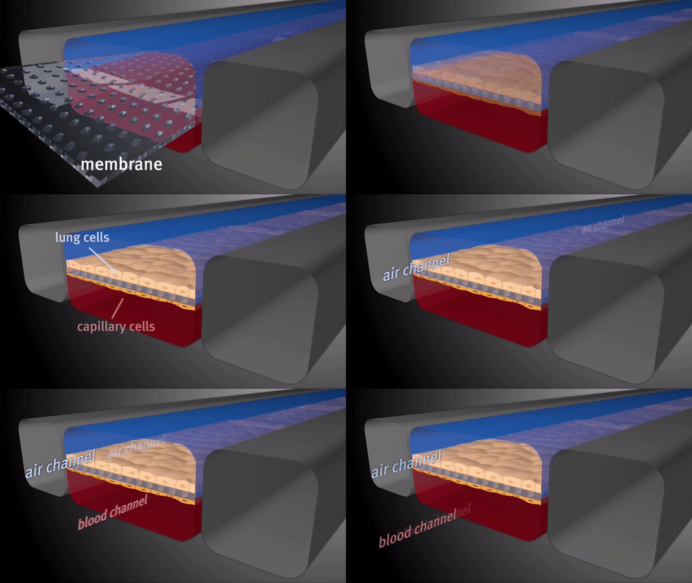
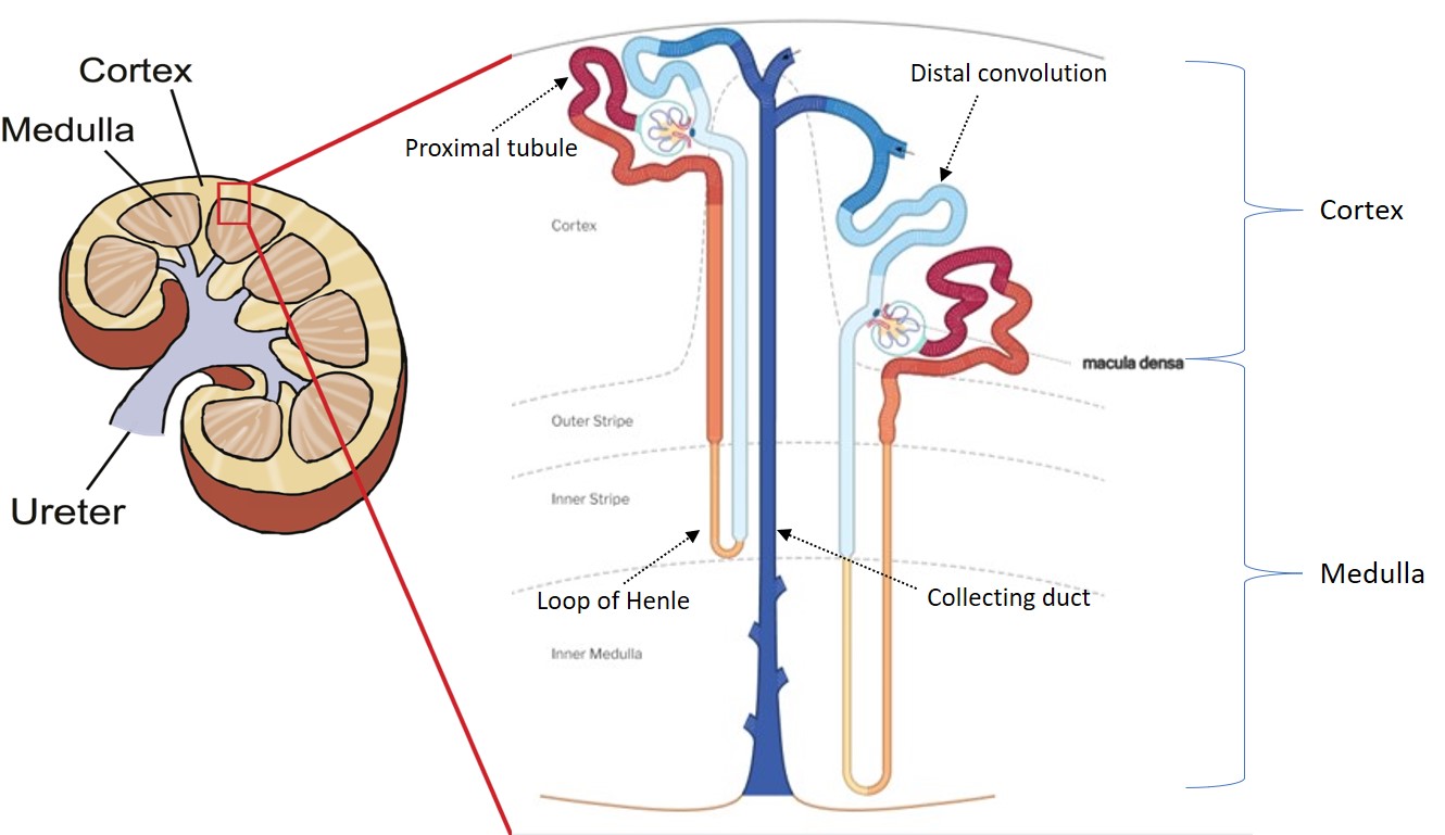
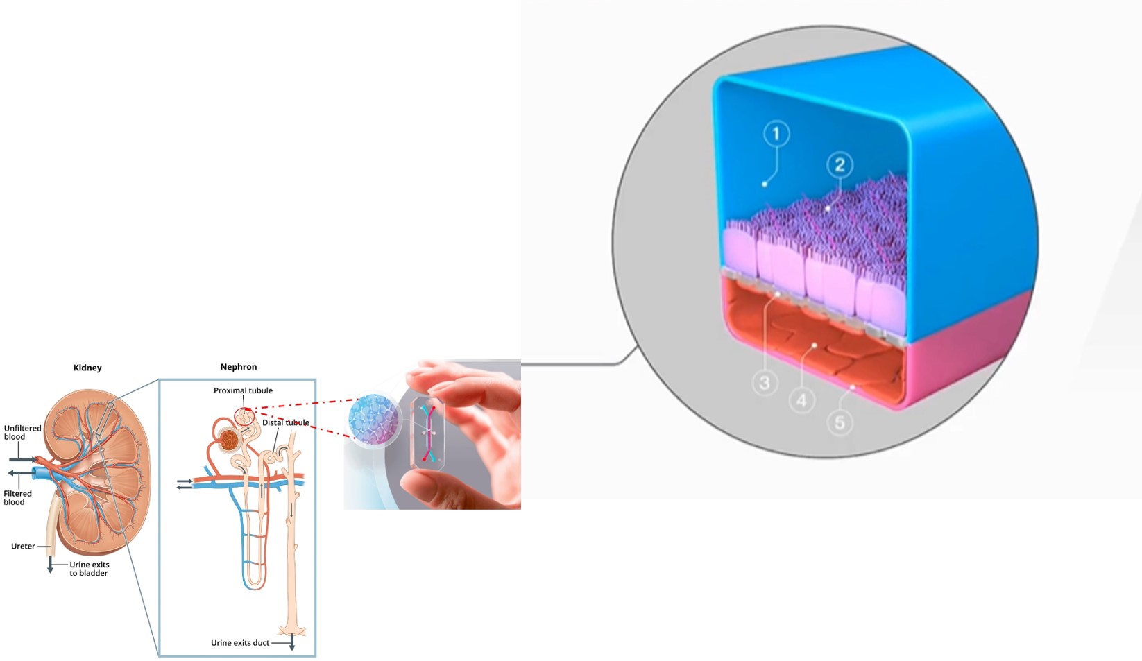
For instance, is it time for reviewer 3 to request human organ-chip experiments instead of animal validation studies? (Ingber D.E. 2020) How effectively can we translate human pathologies within a microenvironment by using patient biopsies in the lab? (Boettner 2018) Can this technology provide us with basic scientific insights at the molecular level to bioengineer a detailed pathological cell signaling pathway underlying disease? And in this way can we guide the clinical development of a personalized, precision diagnostics and therapeutics platform? This post will focus on the kidney, more specifically on the glomerulus and proximal tubule regions of nephrons that can be emulated in the lab on a kidney-chip; an organ-chip instrument that mimics the kidney (Ashammakhi 2018). I am also providing a quick outline on a spectrum of kidney diseases, which include renal biomineralization, chronic kidney disease, and renal carcinomas, closely followed by characterizing the mechanisms of renal tip biomineralization and the role of organ-chips thereon to emulate pathology in vitro.
Hypothesis – emulating pathophysiological stressors underlying renal tip biomineralization on a chip to understand renal disease progression in vitro.
The reference tissue atlas of the human kidney – released by the kidney precision medicine project provides intricate diagrams of the current regions of interest, including the glomerulus and the proximal tubule (Hansen 2022). Several studies have already looked at kidney chip technology in-depth, including those notably conducted at the Wyss Institute for Biologically Inspired Engineering at Harvard, in collaboration with its technological arm Emulate. Inc (Emulate Bio). Of note, early publications chiefly include details behind the microfluidic device-incorporated technology.
- A guide to the microfabrication of human organs-on-chips (Huh 2013, Bhatia 2014)
- Facilitating drug transport and nephrotoxicity assessments on a human kidney proximal tubule-on-a-chip (Jang 2013, Jing 2022), and
- The more recent multi-level organs on a chip, including an intestine-on-a-chip, to illustrate how the recapitulation of human organ-level function at the microscale offers insights on disease pathophysiology, in vitro (Bein 2022).
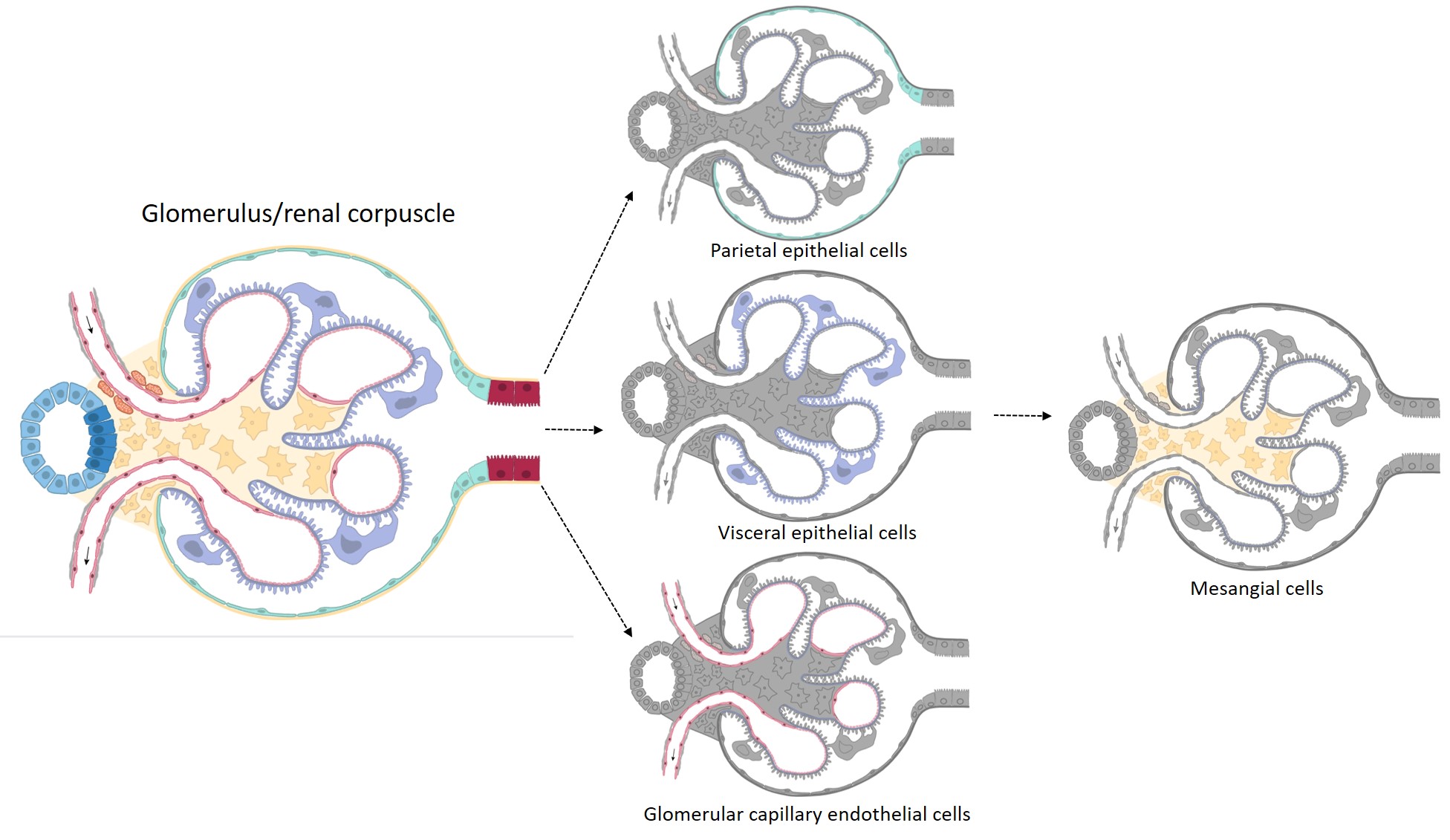
A few variants of organ-on-a-chip models developed to mimic a specific disease model of interest, are listed on table 1.
|
Organ-on-a-chip instrument |
Disease Model |
Reference |
|
A respiring lung-on-a-chip |
A functional alveolar-capillary interface of the human lung |
|
|
Liver-on-a-chip |
A model of fatty liver disease |
|
|
Human kidney proximal tubule-on-a-chip |
For drug transport and nephrotoxicity assessment |
|
|
Reconfigurable multi-organ-on-a-chip |
four, seven or ten different organ models that can interact with one another via microchannels |
|
|
Intestine-on-a-chip |
To mimic nutritional deficiency and the hallmarks of injury of environmental enteric dysfunction |
|
|
Brain-on-a-chip |
Modelling the central nervous system |
Table 1: Variations on a theme. Organ-on-a-chip instruments and the disease models they emulate.
The kidney disease spectrum
In keeping up with the theme of this article, the spectrum of kidney ailments considered here include chronic kidney disease (CKD), renal carcinomas and renal biomineralization, of which chronic diseases affect the structure and function of kidney filtration including nephron tubule portions, to cause a build-up of kidney filter wastes and excess fluids from blood, leading to dangerous levels of fluid, electrolyte and waste buildup in the body (Levey 2012). Co-morbidities of CKD including diabetes, high blood pressure, and cardiovascular disease can impact disease advancement towards end-stage renal disease (MacRae 2021), while some patients also present CKD with unknown etiology (Gashti 2020). Renal tissue mineralization (nephrocalcinosis), stone formation (nephrolithiasis) and Randall’s plaque formation are distinct renal pathologies that contribute to stone formation, and originate in diverse regions of the kidney, with limited information available on the events leading to the initial aggregation of nanometer-scale plaque or stone deposits (Wiener 2019, Ho 2018). Typically stones that form in the renal collecting system are attached to a calcium phosphate lesion of the renal papillae known as Randall’s plaque, while stones can also form in the duct of Bellini, or in free solution (Coe 2010). Renal transcriptomics typically exemplify specific biochemical pathways of interest that contribute to renal disease mechanisms in patients. For example, the hedgehog signaling pathway contributes to renal fibrosis and epithelial mesenchymal transitions (Kramann 2016) during renal biomineralization. Similarly, while renal carcinomas rarely acquire mutations in the p53 tumor suppressor gene (Gurova 2004), clinicopathological outcomes highlight the p53 pathway to be a promising biomarker that predicts the accumulation of a mutant variant, which inhibits the normal functionality of the protein during tumor progression (Wang 2018). Organ-chip instruments provide a proactive platform to investigate key biomarkers of such signaling pathways via gain-of-function or loss-of-function assays under the influence of microphysiological and pathological conditions of stretch, shear-stress and fluid flow.
Pathophysiological stressors underlying renal tip biomineralization, CKD and renal carcinomas
During renal tip biomineralization, increased fluid flow rate that results from co-morbidities of hypertension, hyperfiltration, diabetes, and proteinuria can lead to the build-up of stress to cause chronic shear stress over-time. Increased stimuli of such stressors at the renal tip can hypothetically lead to a localized immuno-modulatory and inflammatory response, characterized by the aggregation of macrophages, the differentiation of cell types including the pathological induction of cellular plasticity, to cause the cellular transition of epithelial mesenchymal stem-cell like phenotypes. These outcomes can lead to fibrosis, apoptosis, cell death, and calcification at the renal tip. As a primary goal, we aim to develop patient-derived, personalized functional assays using renal primary cells, cultured first under static conditions, followed by flow conditions thereafter within organ chip devices. The outcomes of a variety of cell culture techniques can provide insights to predominant biochemical cascades underlying disease progression in a microphysiological environment (Kramann 2016). Moreover, pre-existing data on patient derived renal-tip transcriptomics (RNA-sequencing) have already emphasized the significance of specific biochemical cascades, and their molecular precursors of chronic stress and cell apoptosis, which leads to renal calcification (Fan 2019). The etiology of chronic kidney disease, and renal carcinomas can be characterized similarly on a chip to identify crucial biomarkers of disease progression, in time, for precision medicine and diagnostics applications.
Organ-chip devices – Outlook (and Questions)
In the scenarios described herein, the organ-on-a-chip instrument is a bridge that connects basic science with translational and applied biology. In my opinion, it provides a microphysiological window that can be regulated by the investigator throughout the course of an experiment to observe a desired outcome – to ask and answer fundamental questions of renal disease progression.
- For example, in the context of precision medicine, can oxidative stress at the renal tip be attenuated during progressive renal biomineralization by administering a therapeutic dose of a small molecule drug, after initial nephrotoxicity tests on an organ-chip platform?
- In the context of precision diagnostics, can we identify the fundamental pathological biomarkers of early onset renal biomineralization and early onset chronic kidney disease via patient biopsies cultured on a kidney-chip, to provide a standardized dietary and health guideline for optimized renal health in the long run?
- In a purely basic science approach, can we introduce shear-stress and optimized flow conditions and pathologically relevant stretch parameters on the organ-chip instrument, to mechanically induce and observe the onset of cellular plasticity transitions from a healthy to a pathological state?
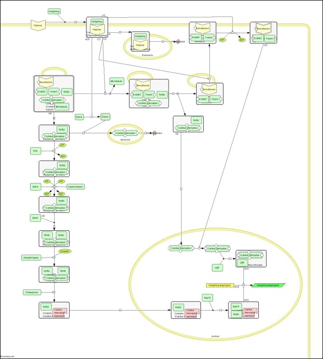
- In this way, in a translational context can we bring together all relevant facets to bottom-up engineer a pathological cell signaling pathway on an organ-chip to identify if a target switch underlies the regulation of renal biomineralization?
- In the context of the interdisciplinary to cross-disciplinary pipeline - based on these findings, can we conduct cross-disciplinary studies in renal oncology, to identify the master players of renal carcinomas in a subset of oncology patients who contribute to the spectrum of kidney disease?
What’s next? (Answers)
The primary aim is to design a series of experiments to translate static culture experiments to an organ-chip microenvironment under flow and stretch conditions. The fundamental hypothesis is that it is possible to recreate key microphysiological transitions at the cellular level during the progression of pathological disease by developing functional assays on a chip, to mimic vital biochemical cascades underlying biomineralization at the renal tip. These outcomes will provide robust insights across interdisciplinary research to investigate if a biochemical switch actively underlies the progression of renal biomineralization, while in parallel allowing us to translate the findings from the lab to the clinic to form a new organ-on-a-chip platform for precision medicine and diagnostics studies.
References.
- Bhatia S.N., Ingber D.E, Microfluidic organs-on-chips, Nature Biotechnology, doi: https://doi.org/10.1038/nbt.2989
- Ingber D.E., Is it Time for Reviewer 3 to Request Human Organ Chip Experiments Instead of Animal Validation Studies? Advanced Science, doi: https://doi.org/10.1002/advs.202002030
- Boettner B. True to type: From human biopsy to complex gut physiology on a chip, website: https://wyss.harvard.edu/news/true-to-type-from-human-biopsy-to-complex-gut-physiology-on-a-chip/
- Ashammakhi N. et al. Kidney-on-a-chip: untapped opportunities, Kidney International, doi: 10.1016/j.kint.2018.06.034.
- Hansen Jens et al. A reference tissue atlas for the human kidney, Science Advances, doi: 10.1126/sciadv.abn4965
- Emulate – Unravel the complexities of human biology. Website: https://emulatebio.com/
- Huh D. et al. Microfabrication of human organs-on-chips, Nature Protocol, doi: 10.1038/nprot.2013.137
- Jang K. et al. Human kidney proximal tubule-on-a-chip for drug transport and nephrotoxicity assessment, Integrative Biology, doi: 10.1039/c3ib40049b
- Jing B et al. Functional Evaluation and Nephrotoxicity Assessment of Human Renal Proximal Tubule Cells on a Chip, Biosensors, doi: 10.3390/bios12090718
- Bein A. et al. Nutritional deficiency in an intestine-on-a-chip recapitulates injury hallmarks associated with environmental enteric dysfunction, Nature Biomedical Engineering, https://doi.org/10.1038/s41551-022-00899-x
- Huh D. et al. Reconstituting organ-level lung functions on a chip, Science, 10.1126/science.1188302
- Hassan S. et al. Liver-on-a-Chip Models of Fatty Liver Disease, Hepatology, doi: 10.1002/hep.31106
- Edington C.D. et al. Interconnected Microphysiological Systems for Quantitative Biology and Pharmacology Studies, Scientific Reports, https://doi.org/10.1038/s41598-018-22749-0
- Maoz B.M. Brain-on-a-Chip: Characterizing the next generation of advanced in vitro platforms for modeling the central nervous system, APL Bioengineering, https://doi.org/10.1063/5.0055812
- Levey A.S., Coresh J. Chronic kidney disease, Lancet, 10.1016/S0140-6736(11)60178-5
- MacRae C. et al. Comorbidity in chronic kidney disease: a large cross-sectional study of prevalence in Scottish primary care, British Journal of General Practice, https://doi.org/10.3399/bjgp20X714125
- Gashti C.N., et al. The Renal Biopsy in Chronic Kidney Disease, Chronic Renal Disease (Second Edition), doi: https://doi.org/10.1016/B978-0-12-815876-0.00073-5
- Wiener S.V. et al. Novel Insights into Renal Mineralization and Stone Formation through Advanced Imaging Modalities, Connective Tissue Research, 10.1080/03008207.2017.1409219
- Ho S.P. et al. Architecture-Guided Fluid Flow Directs Renal Biomineralization, Scientific Reports, doi: https://doi.org/10.1038/s41598-018-30717-x
- Coe F.L. et al. Three pathways for human kidney stone formation, Urology Research, doi: https://doi.org/10.1007/s00240-010-0271-8
- Kramann R. Hedgehog Gli signalling in kidney fibrosis, Nephrology Dialysis Transplantation, https://doi.org/10.1093/ndt/gfw102
- Gurova K.V. et al. p53 pathway in renal cell carcinoma is repressed by a dominant mechanism, Cancer Research, doi: 10.1158/0008-5472.can-03-1541
- Wang Z. et al. Prognostic and clinicopathological value of p53 expression in renal cell carcinoma: a meta-analysis, Oncotarget, doi: 10.18632/oncotarget.21971
- Fan Y. et al. Comparison of Kidney Transcriptomic Profiles of Early and Advanced Diabetic Nephropathy Reveals Potential New Mechanisms for Disease Progression, Diabetes, 10.2337/db19-0204
- Mi H. et al. Large-scale gene function analysis with the PANTHER classification system, Nature Protocol, doi: https://doi.org/10.1038/nprot.2013.092




Please sign in or register for FREE
If you are a registered user on Research Communities by Springer Nature, please sign in