Decoding Brain Tumors: Unleashing the Power of Deep Learning to Grade Gliomas with Precision and Insightful Tumor Microenvironment Analysis what did we do
Published in Protocols & Methods, Computational Sciences, and General & Internal Medicine
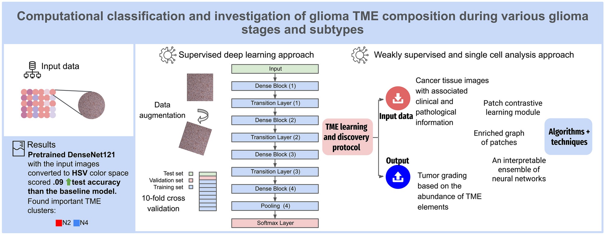
Gliomas, primary brain tumors originating from neural stem cells or glial precursors, pose diagnostic challenges addressed by innovative methodologies. Employing deep learning for automated multiclass tumor grading, our study introduces a protocol exploring tumor microenvironment elements. Despite dataset constraints, image augmentation mitigated imbalances, enhancing accuracy by 9%. The DenseNet121 architecture outperformed, reaching 69%, particularly in WHO grade 2 and 3 cases.

Microenvironment analysis underscored myeloid cells' significance, offering potential diagnostic enhancements for intraoperative evaluations and treatment selection, streamlining workflows for pathologists and oncologists.
In the picture above you can see different tumor grades. However, it is not always so easy and even pathologists have difficulties.
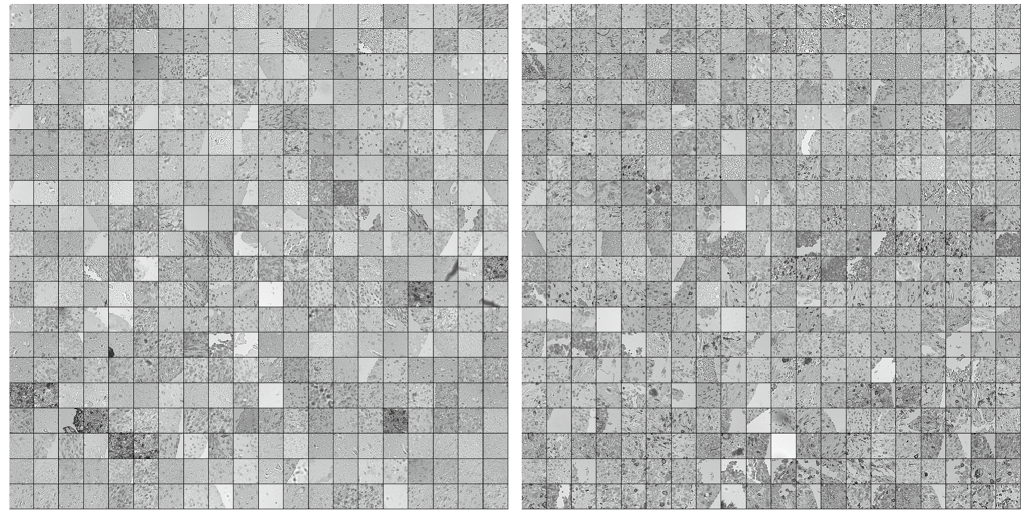
Moreover, if for a second we put aside the classification purpose, and we look carefully what make us differentiating patches of slides of higher grades we found something related to the myeling cells, those neighborhoods are shown below:
Code and used data are available here:
https://github.com/octpsmon/TME_analysis_protocol_n_glioma_grading
Follow the Topic
-
Journal of Imaging Informatics in Medicine
This journal enhances the exchange of knowledge encompassed by the general topic of Imaging Informatics in Medicine including, but not limited to, research and practice in clinical, engineering, information technologies and techniques in all medical imaging environments.

Please sign in or register for FREE
If you are a registered user on Research Communities by Springer Nature, please sign in