Decoding Criminal Behavior: Brain Neurochemical-monitoring Technologies Can Help in Forensic Science
Published in Social Sciences, Protocols & Methods, and General & Internal Medicine
Explore the Research
View of UNDERSTANDING BEHAVIOR PATTERNS IN FORENSIC SCIENCE USING NEUROIMAGING TECHNIQUES
As criminality continues to surge, there's an urgent need for innovative techniques to understand behavioral patterns. The integration of advanced brain imaging technologies, including magnetic resonance imaging, functional magnetic resonance imaging (fMRI), magnetic resonance spectroscopy (MRI), single photon emission computed tomography (SPECT), and positron emission tomography (PET), has played a pivotal role in elucidating structural and functional alterations within brain regions implicated in aggressive or violent behavior, decision-making processes, self-control mechanisms, and reward-seeking conduct. By combining these sophisticated imaging technologies with psychological assessments, researchers have effectively compared individuals displaying heightened levels of violent or aggressive tendencies to those exhibiting less aggressive behavior. Additionally, investigations have delved into comprehending the neural attributes of individuals grappling with brain disorders such as psychotic illnesses, schizophrenia, and antisocial personality disorder.
This interdisciplinary approach holds particular significance in unveiling the intricate connections between brain function and behavior, shedding light on the factors that may contribute to aggressive conduct. In severe instances, these aggressive tendencies may escalate to criminal acts, impacting both individuals with healthy mental health and those affected by disorders, thereby inflicting substantial harm upon victims and society at large.
The exploration of neurobiological factors for the understanding the cause behind criminality and antisocial behavior has witnessed exponential growth in research. Brain regions implicated in decision-making, self-control, reward-seeking, and aggressive/violent conduct include the prefrontal cortex (PFC) comprising the orbitofrontal cortex (OFC), dorsolateral prefrontal cortex (DLPFC), ventromedial prefrontal cortex (VMPFC), as well as the amygdala, striatum, and anterior cingulate cortex (ACC). Investigating these brain regions for structural and functional abnormalities in individuals involved in criminal activities can yield valuable insights for distinguishing and defining their behavior.
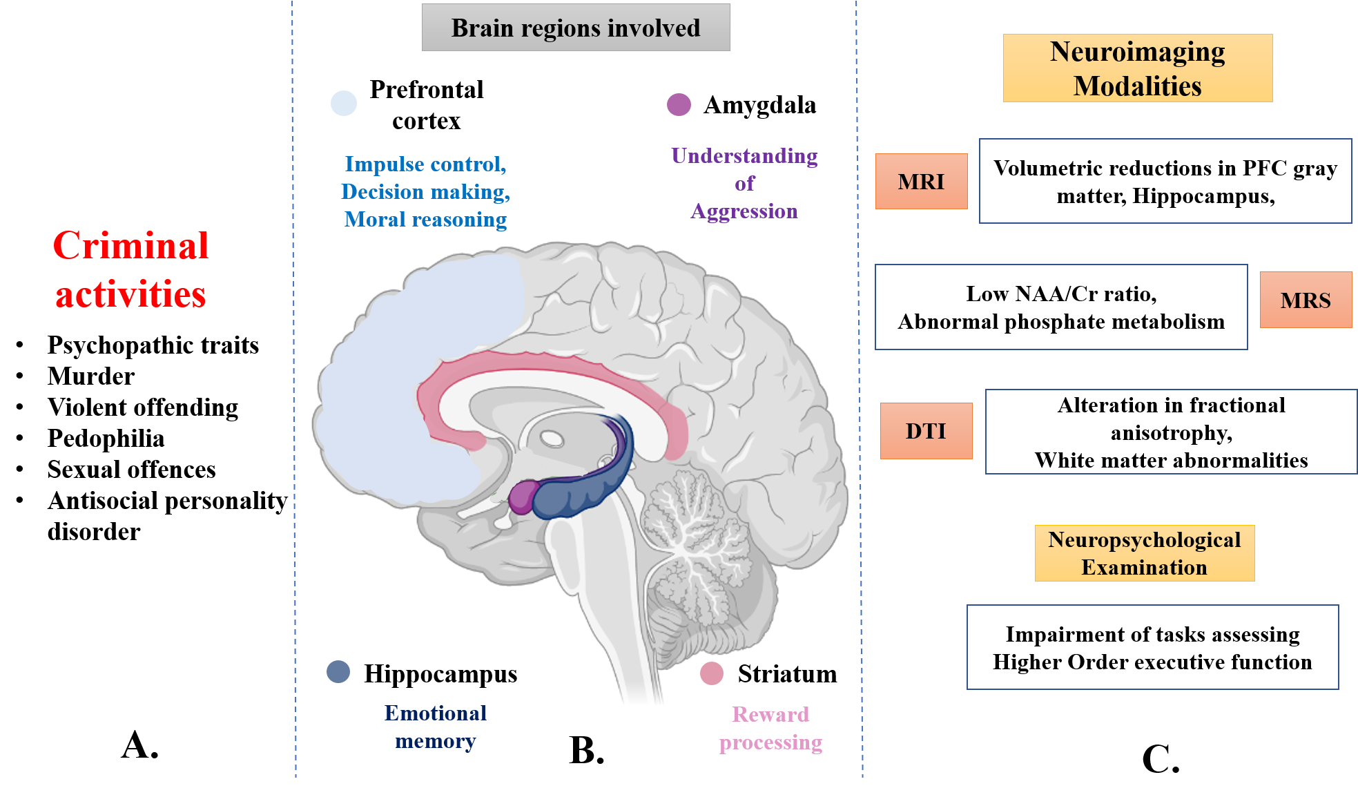
Figure 1: Schematic representation of the association of brain and crime. (A) Criminal activities are witnessed in society. (B) Regions of the brain that are responsible for various functions including impulse control, decision making, emotional memory, reward processing, etc. (C) Observations from different neuroimaging and neuropsychological studies in criminals.
Figure 1(B) was created using Biorender.
Recent advancements in brain imaging techniques have opened new avenues for understanding the neurobiological factors behind criminal behavior. Brain imaging methods like SPECT, PET, fMRI, and MRI have revealed structural and functional anomalies in the brains of individuals with neurological conditions. PET studies show irregularities in brain glucose metabolism, especially in the temporal and prefrontal regions, of antisocial subjects. SPECT has found reduced blood flow in areas like the temporal cortex, prefrontal cortex, and hippocampus in psychiatric patients. The addition of fMRI, which measures the blood-oxygen-level-dependent (BOLD) signal during emotional or cognitive tasks, enables real-time task capture and shows abnormal activation in the hippocampus, amygdala, and frontal regions. Unlike PET and SPECT, fMRI is preferred for its non-invasive nature and real-time task capture. MRI is often used for structural analysis, with studies identifying reduced gray matter volume in the temporal and prefrontal regions of antisocial individuals. Research is also exploring the structural connections between brain regions linked to criminal behavior using diffusion tensor imaging (DTI) to assess white matter microstructure.
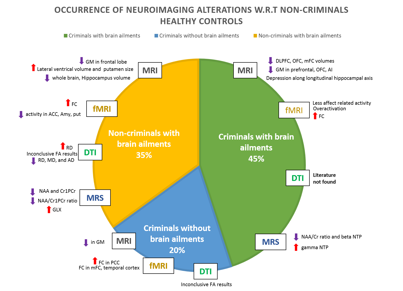
Figure 2: Patterns of neuroimaging outcomes from the three distinct categories i.e. non-criminals with brain ailments, criminals with brain ailments, and criminals without brain ailments. Abbreviations: GM: grey matter; FC: functional connectivity; ACC: anterior cingulate cortex; Amy: amygdala; Put: putamen; RD: radial diffusivity; FA: fractional anisotropy; MD: mean diffusivity; AD: axial diffusivity; NAA N-acetyl-aspartate; Gly: glycine; PCC: posterior cingulate cortex; mFC: medial frontal cortex; DLPFC: dorsolateral prefrontal cortex; OFC: orbitofrontal cortex; MRI: magnetic resonance imaging; MRS: magnetic resonance spectroscopy; fMRI: functional magnetic resonance imaging; DTI: diffusion tensor imaging
Diagrammatic presentation (Figure 2) shows changes in both the structure and function of the brains of individuals engaged in criminal behavior, suggesting a potential link to criminal acts. Addressing these brain region alterations early may help prevent such behavior. However, a definitive conclusion requires further investigation in this field. Given the potential of neuroimaging techniques, there is a thrust for their integration into criminal profiling and investigative practices. This, however, requires careful planning and validation. Ensuring reliability and applicability depends on achieving high levels of sensitivity and specificity, which are essential for acceptance.
Both of us believe that this blog will stimulate this area of research in the coming days with well-predicted features that will help to identify criminals non-invasively and much early before the crime is committed.


Please sign in or register for FREE
If you are a registered user on Research Communities by Springer Nature, please sign in