

Probably the first thing that comes to mind when thinking about biofilms is cell adhesion. A pathogen or potentially pathogenic cell needs to interact with the surface of a host as a key early step in infection. Sometimes this interaction is highly specific, and receptors have been identified on microbial cells and the host that mediate attachment. After this critical step, microbial cells begin to grow and stick together, building on their primary foothold to develop into a biofilm. The importance of these adhesion steps is exemplified by the fact that biofilms are necessary for infection in many microbial species.
The picture of cell adhesion in fungal pathogens is not as clear as one might think. In particular, our knowledge of fungal biofilm formation is lacking in several places. One problem currently faced is that there are many fungal adhesion molecules. Instead of one key adhesion protein, like in Baker’s yeast and several highly studied bacterial pathogens, in some fungal species, like the major opportunistic pathogen Candida albicans there are entire families of related proteins that contribute to adhesion functions. It can be difficult decipher the unique contribution of each adhesion molecule in a biofilm, especially because members in a family can have overlapping or redundant functions. A second challenge in studying fungal biofilms is that the environment of the biofilm is not constant. Cells in a biofilm express different adhesion molecules at different times and in different parts of the biofilm. Sometimes a cell will stop expressing adhesion molecules altogether and fall off the biofilm (become planktonic). Other times, cells express a unique adhesion molecule at a specific time. It remains pretty murky what these sticky molecules are doing during biofilm development, and who is doing what in the correct spatial and temporal context. Moreover, unlike what is seen in the lab, biofilms form in open cavities, like oral tissues. In this complex environment, cells must work against the flow of saliva that continually acts to remove and clean microbes from the mouth. A third challenge is that fungal cells typically change their shape in a biofilm, in a process where they become elongated and twisted in filaments or hyphae. Filamentous/hyphal growth can help the biofilm form in the right pattern, but this process complicates our ability to separate out adhesion functions from other aspects of biofilm development.
In our paper, we addressed these problems in several ways. One was to use flow chambers to mimic the environment of oral tissues and thereby improve studies over normal lab conditions. In particular, by the use of a flow system combined with real-time imaging of C. albicans cells, we were able to watch cells adhere to and grow on surfaces. What we found was pretty surprising. Cell adhesion under flow was a multi-phase process. It was initiated with cells rolling on surfaces to get traction and was followed by firm attachment to the substrate. After attachment, cells entered a growth phase where they either committed either to adherence (adhesion maintenance) or detachment for dispersal to favor planktonic growth.
To define how the different aspects of adhesion were regulated by the biofilm under flow, we took a genetic approach. By examining the behavior of mutants, we found that some proteins were mainly involved in the initial attachment (ALS1/ALS3), while others were required for both initial attachment and prolonged adhesion (EAP1, HWP2, HYR1, and IHD1), which we refer to as adhesion maintenance. Because adhesion maintenance is less well understood than attachment, we explored this property of biofilm development in more detail. We found that the rate of C. albicans detachment was most the important aspect in determining the density of the mature biomass. We also found that hyper-filamantous strains improved total biofilm growth by reducing the rate of detachment. These results demonstrate that hyphal morphogenesis is important for adhesion maintenance in the developing biofilm.
Another way we approached this problem was by a heterologous expression approach. We took the Candida albicans adhesion protein, Ywp1, which we suspected to mainly function in adhesion maintenance (the protein was previously thought to regulate dispersion) and expressed it in the Baker’s yeast Saccharomyces cerevisiae, which has less adhesion molecules and is relatively easy to manipulate, but that nevertheless makes biofilms and undergoes filamentous growth. We then asked what does Ywp1 do as a stand-alone adhesion molecule? Technically, the experiment was challenging, because the codon preference between the two species is different. We took a synthetic biology approach to design and express a tagged version of C. albicans Ywp1 in S. cerevisiae. This experiment validated the flow results in C. albicans, demonstrating that Ywp1 may be the first example of an adhesion maintenance protein in C. albicans. Collectively, the study helps get us one step closer to understanding the real-life world of fungal biofilm adhesion regulation.
Follow the Topic
-
npj Biofilms and Microbiomes

The aim of this journal is to serve as a comprehensive platform to promote biofilms and microbiomes research across a wide spectrum of scientific disciplines.
Related Collections
With Collections, you can get published faster and increase your visibility.
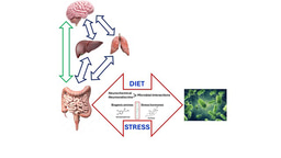
Natural bioactives, Gut microbiome, and human metabolism
Publishing Model: Open Access
Deadline: Feb 20, 2026
Harnessing plant microbiomes to improve performance and mechanistic understanding
Publishing Model: Open Access
Deadline: Jun 01, 2026


Please sign in or register for FREE
If you are a registered user on Research Communities by Springer Nature, please sign in
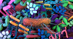
Love your poster image!