Finding gold in silver-stained rat brains
Published in Neuroscience and Research Data

Throughout history, neuroscientists have studied and used brain material from small animals to carry out experiments to better understand the structure of the brain. In recent years, technology has made reuse more relevant in brain research. If we can utilize and reuse research material we already have, and thus reduce future animal experiments, we will be able to save resources on several levels.
Historical collection of rat brains
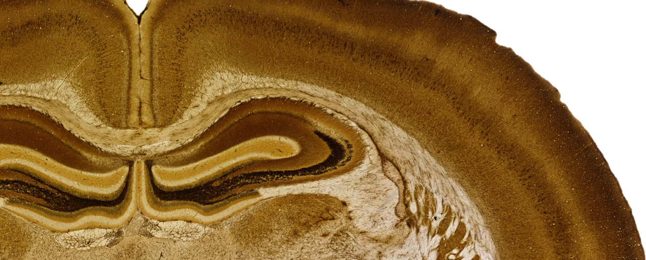
During the 1970s, a significant collection of rat brains was treated with silver staining to study and map the normal rat brain. The historical collections have been hidden on dusty shelves for several decades. This sulphide silver staining, originally developed by Friedrich Timm and later modified by Finn-Mogens Šmejda Haug, reveal neuronal perikarya, neuropil and glial processes under a microscope, thus making it possible to distinguish between different areas of the brain. The brain sections were neatly placed on microscope slides and were described in several articles1-3 published from 1973-76 by Haug and colleagues. These articles contained a few sample images and otherwise detailed descriptions of what could be seen in the microscope. A group of brain researchers at the University of Oslo wanted to change the fate of the silver rat material. It turns out that the rat brains can be of great use to students and researchers who want to understand the normal rat brain and study brain disorders.
Sharing a collection of rat brain data
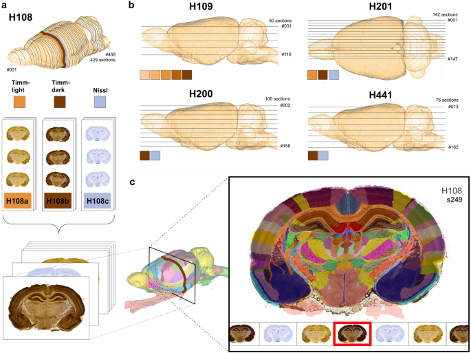
Today, neuroscientists have good opportunities to share more than what the format of a paper publication allows. Together with data curation scientists from the Human Brain Project and EBRAINS, the complete rat brain collection has been made available so that others outside our laboratory can see entire series of brain sections from the normal rat brain. The rat material consists of five brains, where each brain was frozen and cut into at least 150 sections. We have scanned and digitized 797 brain sections, which have turned into 797 images. As the brains are cut in different orthogonal planes, one coronal, one sagittal and three horizontal, looking at the entire series of brain sections from one end to the other may help neuroscientists to gain a three-dimensional understanding of the brain's organization.
Web microscope and atlas of the rat brain
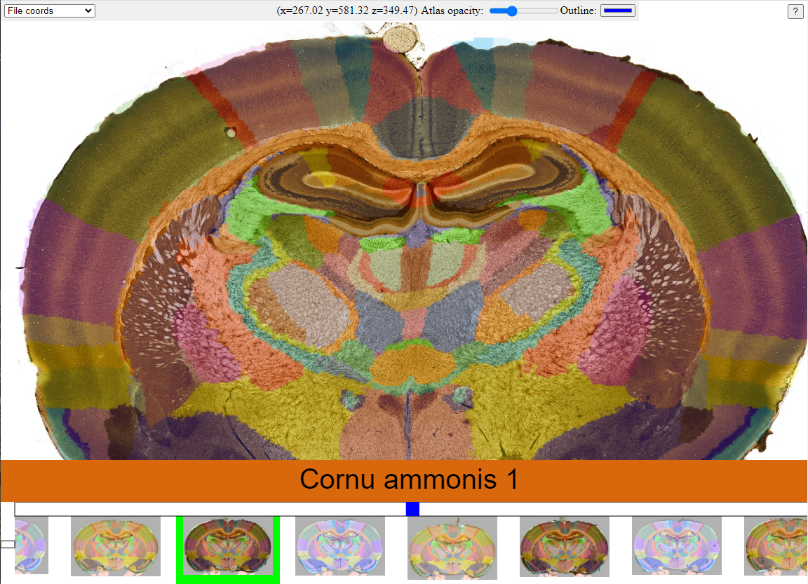
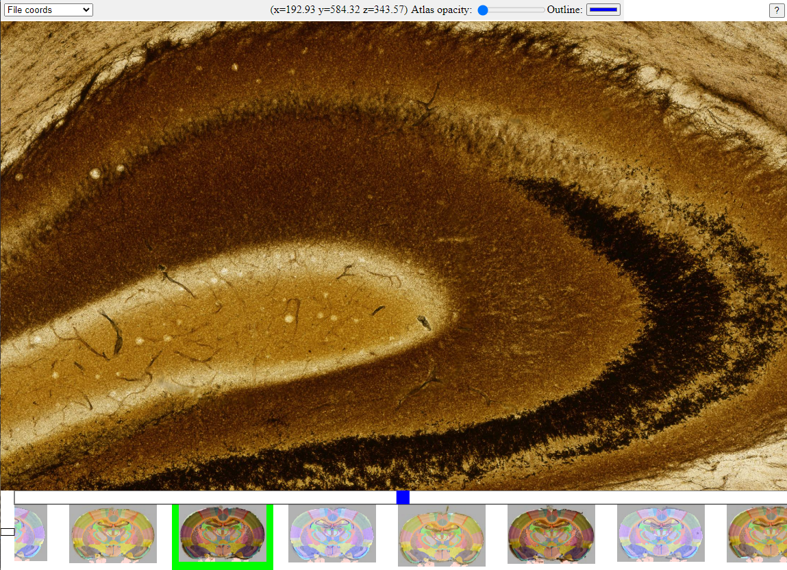
Each of the digitized rat brain section images have such a high resolution that you can zoom in and study the tissue as you would do with an analogue microscope. It is therefore appropriate that all the images are placed in a web microscope. With this application, one can study a series of brain sections from the same rat brain in sequential order. This gives the opportunity to follow a brain area through several brain sections. With the help of a 3D atlas, the Waxholm Space rat brain atlas4-5 (v4; RRID: SCR_017124), lines are drawn along different brain areas inside the virtual microscope, so that it becomes easier to distinguish between neighboring areas and study any differences.


Screenshots of the web microscope.
Anyone can study the brain
The web microscope is openly available to anyone via EBRAINS.eu (https://search.kg.ebrains.eu), without login or registration. To go to the rat brain collection, you can use the DOI links in the related data publications6-7. Both students, researchers and curious others now have a lot of material from the normal rat brain they can look through. The data collection is suitable as a benchmark reference of the normal rat brain, for planning new studies in general or specifically for further investigations of zinc-related phenomena. This data sharing effort also exemplifies how valuable historical histological collections can be revived and made accessible for future research.
References
1 Haug, F. M. Š. Heavy metals in the brain. A light microscope study of the rat with Timm's sulphide silver method. Methodological considerations and cytological and regional staining patterns. Adv. Anat. Embryol. Cell Biol. 47, 1-71 (1973).
2 Haug, F. M. Š. Light microscopical mapping of the hippocampal region, the pyriform cortex and the corticomedial amygdaloid nuclei of the rat with Timm's sulphide silver method. I. Area dentata, hippocampus and subiculum. Z. Anat. Entwicklungsgesch. 145, 1-27 (1974).
3 Haug, F. M. Š. Sulphide silver pattern and cytoarchitectonics of parahippocampal areas in the rat. Special reference to the subdivision of area entorhinalis (area 28) and its demarcation from the pyriform cortex. Adv. Anat. Embryol. Cell Biol. 52, 3-73 (1976).
4 Papp, E. A., Leergaard, T. B., Calabrese, E., Johnson, G. A. & Bjaalie, J. G. Waxholm Space atlas of the Sprague Dawley rat brain. NeuroImage. 97, 374-386 (2014).
5 Kleven, H., Bjerke, I.E., Clascá, F. et al. Waxholm Space atlas of the rat brain: a 3D atlas supporting data analysis and integration. Nat Methods 20, 1822–1829. https://doi.org/10.1038/s41592-023-02034-3 (2023).
6 Blixhavn, C. H. et al. Multiplane microscopic atlas of rat brain zincergic terminal fields and metal-containing glia stained with Timm's sulphide silver method (v1). EBRAINS https://doi.org/10.25493/T686-7BX (2022).
7 Blixhavn, C. H. et al. Contrast reference images for the Timm-Haug73 modification of Timm's sulphide silver method (v1). EBRAINS https://doi.org/10.25493/4K9X-FJW (2022).
Follow the Topic
-
Scientific Data

A peer-reviewed, open-access journal for descriptions of datasets, and research that advances the sharing and reuse of scientific data.
Your space to connect: The Psychedelics Hub
A new Communities’ space to connect, collaborate, and explore research on Psychotherapy, Clinical Psychology, and Neuroscience!
Continue reading announcementRelated Collections
With Collections, you can get published faster and increase your visibility.
Data for crop management
Publishing Model: Open Access
Deadline: Apr 17, 2026
Genomics in freshwater and marine science
Publishing Model: Open Access
Deadline: Jul 23, 2026


Please sign in or register for FREE
If you are a registered user on Research Communities by Springer Nature, please sign in