Harnessing red blood cell vesicles for gene therapies
Published in Bioengineering & Biotechnology

The paper in Nature Communications is here: https://go.nature.com/2tnLfMn
Many people who work on extracellular vesicles (EVs) have found that it is too labor-intensive to purify sufficient EVs for functional assays. I knew this all too well from my postdoctoral training at Harvard Medical School when I had to work with liters, not milliliters, of conditioned media, over long hours of cell culture and ultracentrifugation every day.1
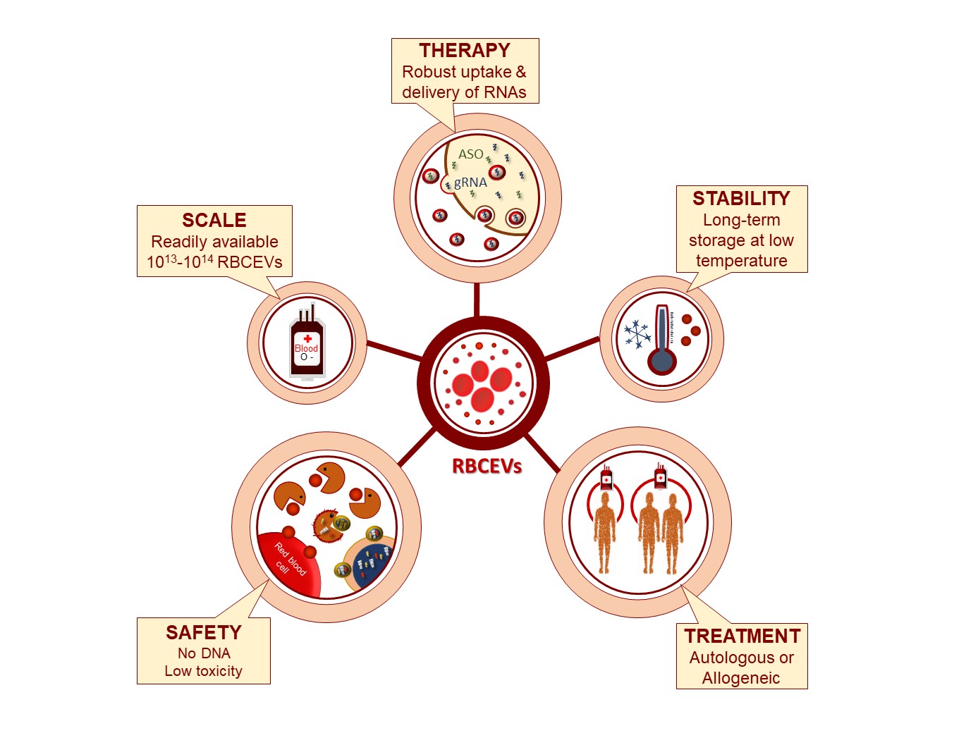
When I started my own new lab at the City University of Hong Kong (CityU) in Aug 2015, one of our projects was to target miR-125b in acute myeloid leukemia (AML) using antisense oligonucleotides (ASOs) in lipid nanoparticles, based on my previous studies on miR-125b.2,3 Unfortunately, we found that the nanoparticles did not work in leukemia cells although they worked very well in other cell types (in a collaboration with Prof. Michael Yang’s and Dr. Linfeng Huang’s labs at CityU). So we thought of trying EVs because some previous studies had shown that EVs are especially good delivery vehicles for blood cells.4 But herein lies the problem: how do we get enough EVs for delivering the ASOs? It would be ideal if we could use a readily available source of blood cells, such as red blood cells (RBCs). I recalled what I learned 12 years ago in the Lodish lab about RBCs and discussed extensively with my colleague Dr. Jiahai Shi, who worked on novel RBC therapies at CityU, about using RBCs for EV production. In May 2016, for the first time, we purified red blood cells derived EVs (RBCEVs) using ultracentrifugation and found an amazingly thick layer of red EVs on a sucrose cushion from only 50 ml of RBCs. After washing carefully, we still got more than 1013 RBCEVs, a scale that we had never archived from other cell types. We electroporated RBCEVs with fluorescent ASOs, then exposed AML cells to these EVs. To our surprise, almost 80% of the AML cells became fluorescence positive!
Our project progressed very quickly after Waqas Usman joined my lab as a PhD student in Sep 2016 and we started a good collaboration with Dr. William Cho’s group who provided us great help and resources. Worryingly, Waqas found that EVs derived from the whole blood provided by Red Cross led to an increase in leukemia cell proliferation. This could be due to proto-oncogenic or growth-stimulating factors in the leucocytes and lymphocytes. Thus, he separated RBCs completely from other cell types and from the plasma to avoid this worrying effect. It worked, and we obtained very high yields of pure EVs from RBCs. We further optimized the ASO electroporation procedure and obtained very efficient (80-90%) knockdown of miR-125b. At the same time, Dr. Jiahai Shi suggested the idea of CRISPR/Cas9 delivery via RBCEVs and worked closely with us on this experiment. We were unsure if the Cas9 mRNA was too large for RBCEVs initially, but it turned out well after some optimization. We were amazed that RBCEVs had such an impressive therapeutic potential hence, we quickly moved forward with the manuscript writing and submission.
During the manuscript revision, the most critical question that was raised was whether RBCEVs could deliver the ASOs in a systemic manner. We learned to use NSG mice for AML engraftment with help from Prof. Anskar Leung’s lab. We found that i.p. injection of the RBCEVs got them very quickly into the liver, spleen and bone marrow where leukemia typically develops. So, we treated the leukemic mice with a high dose of anti-miR-125b RBCEVs via i.p. injection every other day. Remarkably, the treatments showed a good therapeutic effect in only 9 days.
For us, the most remarkable feature of the RBCEV platform is the ease of getting the EVs in large quantities without any expensive and labor-intensive culture. The beauty of this platform is the robust delivery to many cell types, including leukemia cells, and the safe contents of RBCEVs, which are devoid of DNA, growth factors, or potentially immunogenic and toxic substances because RBCs are enucleated ghost cells. We are currently working on further characterization and modification of RBCEVs so that we can completely weaponize the platform for cancer-specific delivery of gene therapies.

Original article: Usman et al, Efficient RNA drug delivery using red blood cell extracellular vesicles, Nature Communications, volume 9, Article number: 2359 (2018).
References
1. Le, M. T. N. et al. miR-200–containing extracellular vesicles promote breast cancer cell metastasis. J. Clin. Invest. 124, 5109–5128 (2014).
2. Le, M. T. N. et al. MicroRNA-125b is a novel negative regulator of p53. Genes Dev. 23, 862–876 (2009).
3. Le, M. T. N. et al. Conserved regulation of p53 network dosage by microRNA-125b occurs through evolving miRNA-target gene pairs. PLoS Genet. 7, e1002242 (2011).
4. Wahlgren, J. et al. Plasma exosomes can deliver exogenous short interfering RNA to monocytes and lymphocytes. Nucleic Acids Res. 40, e130 (2012).


Please sign in or register for FREE
If you are a registered user on Research Communities by Springer Nature, please sign in