JMCR: Clinical Reasoning From Case Reports
Published in Neuroscience and Biomedical Research
Explore the Research
Diagnostic dilemma in the management of a cervical spinal cord lesion: neuromyelitis optica spectrum disorder: a case report
Adapted from: Azode KM, Balogun SA, Komolafe MA, Olasode BJ, Komolafe EO. Diagnostic dilemma in the management of a cervical spinal cord lesion: neuromyelitis optica spectrum disorder: a case report. Journal of Medical Case Reports. 2026. doi:10.1186/s13256-026-06420-1.
Introduction
An expansile, enhancing intramedullary cervical spinal cord lesion accompanied by rapidly progressive quadriplegia immediately raises concern for a structural spinal cord process. But cord enlargement does not always mean tumor.
This case demonstrates how longitudinally extensive transverse myelitis, optic nerve abnormalities, vomiting, and rapidly progressive respiratory compromise can redirect the diagnostic reasoning toward neuromyelitis optica spectrum disorder (NMOSD)—and why distinguishing inflammatory cord expansion from surgically treatable compression matters.
Clinical Case
A 50-year-old right-handed Nigerian woman of Yoruba ethnicity presented with progressive weakness involving all four limbs over approximately 2 weeks.
The weakness began around the left shoulder, progressed through the left upper limb, and subsequently involved the remaining extremities until she became quadriplegic.
Approximately 1 month before presentation, she had experienced a week-long febrile illness with sore throat that resolved after antibiotic and antimalarial treatment.
Additional symptoms included photophobia, recurrent vomiting, difficulty swallowing, and later difficulty breathing.
Neurologic examination demonstrated increased tone and hyperreflexia in all four limbs with 0/5 motor power throughout. Motor and sensory levels were reported around C4 and C5, respectively. Joint-position and vibration sensation were absent bilaterally.
Rectal examination demonstrated absent perianal and deep anal pressure sensation, lax anal tone, and an absent bulbocavernosus reflex.
The patient also had lower cranial nerve abnormalities, including impaired deglutition and tongue weakness with deviation to the left.
First Teaching Pause: Define the Syndrome Before Naming the Disease
The first task is localization.
This patient had:
-
Rapidly progressive quadriplegia
-
Diffuse upper motor neuron findings
-
A cervical sensory level
-
Posterior-column sensory loss
-
Sphincter dysfunction
-
Bulbar manifestations
-
Emerging respiratory compromise
The dominant syndrome was therefore a severe high cervical myelopathy.
The relatively rapid progression over approximately 2 weeks should broaden the differential beyond neoplasm. Tumors can certainly produce cord enlargement and severe deficits, but an acute-to-subacute course also raises inflammatory, infectious, vascular, and other non-neoplastic possibilities.
The localization can be summarized as:
A rapidly progressive, severe cervical myelopathy with additional symptoms suggesting possible involvement beyond the spinal cord itself.
That final point becomes important.
Initial Differential Diagnosis
Intramedullary spinal cord neoplasm
The combination of cord expansion and an intramedullary MRI abnormality makes an intramedullary tumor an understandable initial concern.
Potential considerations include ependymoma, astrocytoma, and other primary or secondary spinal cord lesions.
However, imaging morphology must be interpreted together with clinical tempo and lesion length.
Foramen magnum or other high cervical structural lesion
The authors initially considered a high cervical or foramen magnum lesion because of the profound quadriplegia, bulbar findings, and respiratory deterioration.
A structural lesion at this level could plausibly account for severe motor and respiratory dysfunction.
Neuromyelitis optica spectrum disorder
NMOSD becomes increasingly important when a patient has an extensive inflammatory-appearing spinal cord lesion together with findings potentially involving the optic nerve, brainstem, or area postrema.
Myelin oligodendrocyte glycoprotein antibody-associated disease
MOG antibody-associated disease can also produce extensive inflammatory myelitis and was appropriately included in the differential.
Multiple sclerosis
Multiple sclerosis remains part of the differential diagnosis of inflammatory myelopathy, although the pattern of a very long spinal cord lesion should prompt consideration of alternatives.
Other inflammatory, infectious, or vascular myelopathies
The rapid clinical progression requires consideration of additional causes of acute or subacute myelopathy rather than assuming that an expansile MRI appearance necessarily represents tumor.
Imaging and the Diagnostic Pivot
Brain MRI was interpreted by the authors as showing abnormalities involving the right optic nerve.
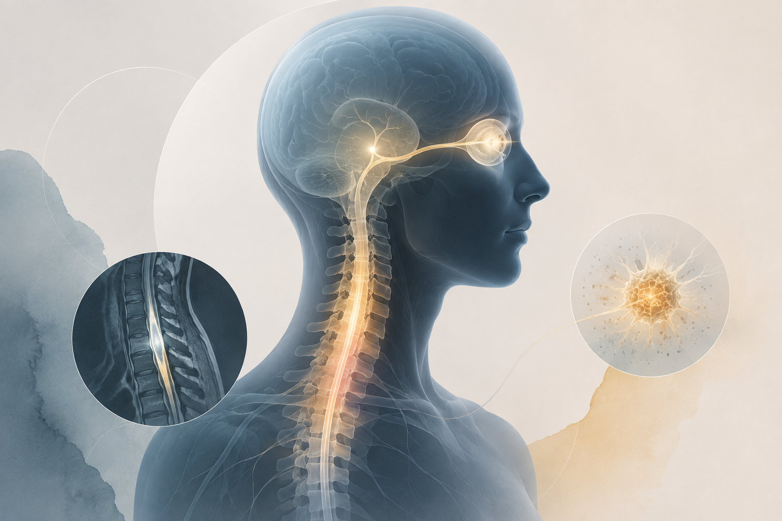
Cervical spine MRI demonstrated a striking intramedullary abnormality with substantial cord expansion and increased T2 signal.
Importantly, the source report describes the longitudinal extent somewhat differently in different portions of the manuscript. The text describes the lesion as extending from approximately C3 to T2, whereas the figure description characterizes prominent bulbous cord expansion and signal abnormality from approximately C3 to C7.
Regardless of the precise reported endpoint, the essential imaging feature was clear:
This was a long-segment intramedullary cervical cord lesion.
Second Teaching Pause: When Does an Intramedullary Lesion Stop Looking Like a Tumor?
The MRI abnormality was expansile.
That observation can create a powerful cognitive anchor:
Expanded cord = mass lesion.
But inflammatory myelitis can also produce dramatic swelling.
The more useful question is not simply:
“Is the cord enlarged?”
It is:
“What disease process best explains the imaging pattern, clinical tempo, and abnormalities elsewhere in the nervous system?”
Several clues now favor an inflammatory disorder:
-
Progression over approximately 2 weeks
-
Long-segment spinal cord involvement
-
Possible optic nerve involvement
-
Recurrent vomiting
-
Bulbar symptoms
-
Rapid respiratory deterioration
The vomiting is particularly interesting. In NMOSD, otherwise unexplained nausea, vomiting, or hiccups can reflect area postrema involvement.
The patient therefore did not simply have an isolated spinal cord lesion. Her symptoms potentially involved several regions characteristically affected in NMOSD.
Clinical Deterioration
On the third hospital day, the patient developed severe respiratory distress and required transfer to the intensive care unit.
Serum testing for aquaporin-4 immunoglobulin G (AQP4-IgG) had been requested, but the assay was not locally available and the specimen had to be sent to another facility.
Meanwhile, the patient's respiratory status continued to deteriorate.
Because of the diagnostic uncertainty and concern for a potentially treatable structural lesion, surgical exploration was pursued.
On hospital day 4, she underwent:
-
C3–C6 laminectomy
-
Intramedullary exploration
-
Biopsy of the spinal cord at approximately C5
Intraoperatively, however, the dura and spinal cord appeared grossly normal, without an obvious intramedullary tumor.
Third Teaching Pause: Swelling Is Not the Same as Compression
This is the central clinical reasoning point in the case.
An enlarged spinal cord caused by intrinsic inflammatory edema is fundamentally different from a spinal cord compressed by an external lesion.
Both may produce:
-
Severe weakness
-
Respiratory compromise
-
Cord enlargement
-
T2 signal abnormality
-
Contrast enhancement
But their pathophysiology—and therefore their treatment—is different.
Before proceeding toward biopsy or decompression of an apparent intramedullary lesion, clinicians should ask:
-
Is there a discrete mass?
-
How rapidly did the syndrome evolve?
-
Is the lesion longitudinally extensive?
-
Are there optic nerve abnormalities?
-
Are there brainstem or area postrema symptoms?
-
Have AQP4-IgG and MOG antibody testing been obtained?
-
Could inflammatory myelitis provide a single explanation for the entire syndrome?
This does not mean that every long spinal cord lesion is inflammatory.
It means that the MRI must not be interpreted independently of the clinical syndrome.
Diagnostic Evaluation
The diagnostic turning point came on the second postoperative day.
Serum testing demonstrated:
-
AQP4-IgG titer: 1:32 — elevated
-
MOG antibody: not elevated
The report also documented markedly elevated C-reactive protein and D-dimer levels.
Histopathologic examination of the biopsied spinal cord tissue showed an inflammatory process without evidence of malignancy. The reported findings included reactive glial cells with ballooning and cytoplasmic clearing, edema, hemorrhage, and capillary proliferation.
The absence of malignancy and the positive AQP4-IgG result substantially reorganized the case.
Evolving Clinical Reasoning
The diagnostic pathway can be reconstructed as a series of probability-changing observations.
Stage 1: Rapidly progressive quadriplegia
The initial syndrome localized to the high cervical spinal cord.
A structural lesion was possible, but the short time course required a broader differential.
Stage 2: Long-segment intramedullary cord abnormality
MRI demonstrated substantial spinal cord expansion and T2 signal abnormality.
The expansile appearance increased concern for an intramedullary lesion, but the longitudinal extent should also raise inflammatory myelopathy.
Stage 3: Symptoms beyond the spinal cord
Vomiting, swallowing difficulty, respiratory dysfunction, and possible optic nerve involvement suggested that the disease might not be confined to a single focal cervical lesion.
Stage 4: Surgical exploration
No obvious intramedullary tumor was identified.
This reduced the probability that the MRI abnormality represented a conventional structural mass.
Stage 5: AQP4-IgG positivity
An elevated serum AQP4-IgG titer provided the key disease-specific laboratory evidence.
Stage 6: Histopathology
The biopsy showed inflammatory neural tissue without malignancy, further supporting an inflammatory rather than neoplastic process.
The individual observations therefore converged on a unifying diagnosis.
Final Diagnosis
Aquaporin-4 antibody-positive neuromyelitis optica spectrum disorder presenting with severe longitudinally extensive cervical myelitis, with possible optic nerve and area postrema involvement.
Diagnostic Discussion
NMOSD is an autoimmune central nervous system disorder associated in many patients with antibodies directed against aquaporin-4, a water channel concentrated in astrocytic end-foot processes.
The source report emphasizes several core clinical manifestations relevant to this case, including:
-
Longitudinally extensive transverse myelitis
-
Optic nerve involvement
-
Area postrema syndrome
-
Brainstem syndromes
-
Respiratory and swallowing difficulties
The spinal MRI appearance created the principal diagnostic dilemma.
Marked inflammatory edema can produce substantial cord expansion and resemble an intramedullary tumor. Conversely, assuming that every long spinal cord lesion is inflammatory would also be unsafe.
The diagnostic reasoning therefore depends on integration.
In this patient, the combination of rapid progression + extensive spinal cord involvement + possible optic nerve abnormality + vomiting + bulbar/respiratory symptoms + AQP4-IgG positivity provided a more coherent explanation than an isolated cervical spinal cord neoplasm.
Management Discussion
Postoperatively, the patient remained in the intensive care unit and required mechanical ventilatory support.
The reported treatment included:
-
Prednisolone
-
Azathioprine
-
Ventilatory support
-
Nasogastric feeding
-
Antimicrobial therapy
-
Anticoagulant therapy
-
Tenofovir in the setting of hepatitis B surface antigen positivity
-
Cardiopulmonary and neurophysiotherapy
A tracheostomy was performed on postoperative day 12.
The authors reported a reduction in respiratory distress after immunosuppressive treatment was started.
The source article also discusses corticosteroids, immunosuppressive therapy, and plasma exchange as approaches used in NMOSD. Plasma exchange, however, was not reported as having been administered to this patient.
This distinction is important: educational discussion of treatment options should not be confused with what actually occurred in the individual case.
Outcome and Follow-Up
Despite intensive care and respiratory support, the clinical course remained severe.
On postoperative day 27, the patient developed sudden respiratory distress followed by hemodynamic instability.
Cardiopulmonary resuscitation was unsuccessful, and she died.
The precise cause of the terminal deterioration was not established.
The authors considered the possibility of disease relapse, but the available report does not definitively demonstrate the mechanism of the final respiratory event.
That uncertainty should be preserved.
CARE Timeline
| Time | Clinical events |
|---|---|
| Approximately 1 month before presentation | One-week febrile illness with sore throat, subsequently resolved. |
| Approximately 2 weeks before admission | Progressive weakness began around the left shoulder and spread until all four limbs were involved. |
| Admission | Quadriplegia, increased tone and reflexes, cervical sensory findings, posterior-column sensory loss, vomiting, swallowing difficulty, and lower cranial nerve abnormalities documented. |
| Initial imaging | Brain MRI interpreted as showing right optic nerve involvement; cervical MRI showed a long-segment expansile intramedullary cord abnormality. |
| Hospital day 3 | Severe respiratory distress developed; patient transferred to intensive care. |
| Hospital day 4 | C3–C6 laminectomy, intramedullary exploration, and spinal cord biopsy performed. No obvious tumor was identified intraoperatively. |
| Postoperative day 2 | Serum AQP4-IgG returned elevated at 1:32; MOG antibody was not elevated. |
| Subsequent course | Histology showed inflammatory neural tissue without malignancy. Prednisolone and azathioprine were among the therapies administered. |
| Postoperative day 12 | Tracheostomy performed. |
| Postoperative day 27 | Sudden respiratory distress and hemodynamic instability developed; resuscitation was unsuccessful. |
Cognitive Pitfalls
Anchoring on the MRI
Cord enlargement and enhancement can immediately suggest tumor.
Inflammatory myelitis, however, can also produce striking cord expansion.
Equating expansion with compression
An intrinsically swollen spinal cord is not necessarily experiencing a surgically reversible compressive process.
Determining why the cord is enlarged matters more than simply documenting that it is enlarged.
Ignoring symptoms outside the spinal cord
Vomiting, possible optic nerve abnormalities, dysphagia, and respiratory dysfunction should not automatically be treated as unrelated problems.
Together, they may identify a multisite inflammatory central nervous system disorder.
Underweighting the clinical tempo
A lesion that produces profound neurologic deterioration over approximately 2 weeks demands consideration of inflammatory and other acute-to-subacute causes even when the MRI resembles a mass.
Waiting for a single test to define the syndrome
AQP4-IgG ultimately provided the decisive laboratory evidence, but several clinical clues were already present.
Diagnostic reasoning should evolve continuously as the history, examination, imaging, and laboratory data accumulate.
Overinterpreting the terminal event
The patient died after sudden respiratory deterioration, but the precise mechanism was not established.
Possible relapse should remain a hypothesis rather than be presented as a demonstrated cause.
Clinical Pearls
-
Long spinal cord lesions require a broad differential. An expansile intramedullary abnormality is not synonymous with neoplasm.
-
Clinical tempo matters. Rapid progression over days to weeks should increase suspicion for inflammatory, infectious, vascular, and other acute myelopathies.
-
Look beyond the cord. Optic nerve, brainstem, and area postrema manifestations may provide the clues that unify an apparently isolated spinal lesion.
-
Vomiting can be neurologic. In the appropriate clinical context, recurrent unexplained vomiting may reflect area postrema involvement in NMOSD.
-
Cord swelling is not the same as mechanical compression. The distinction has major therapeutic consequences.
-
AQP4-IgG can be the diagnostic pivot. Early testing is particularly valuable when longitudinally extensive myelitis is accompanied by other characteristic clinical features.
-
Resource limitations can alter the diagnostic sequence. In this case, AQP4-IgG testing had to be performed at another facility and the result became available only after surgery.
-
Preserve uncertainty. A clinical reasoning discussion should distinguish what the case demonstrates from what remains suspected or unexplained.
Clinical Take-Home Message
An expansile cervical spinal cord lesion should not automatically be classified as a tumor.
In this patient, the rapid progression to quadriplegia, long-segment spinal cord involvement, recurrent vomiting, possible optic nerve abnormality, bulbar and respiratory manifestations, and eventual AQP4-IgG positivity pointed toward NMOSD.
The central lesson is one of diagnostic integration: the morphology of an MRI lesion must be interpreted in the context of clinical tempo, neurologic localization, associated syndromes, and disease-specific testing.
Recognizing that an enlarged spinal cord may represent inflammatory edema rather than a surgically correctable mass can fundamentally change the diagnostic and therapeutic pathway.
Board-Style Questions
Question 1
A 50-year-old woman develops rapidly progressive quadriplegia over 2 weeks. Cervical MRI demonstrates a long-segment expansile intramedullary T2-hyperintense lesion. Which diagnosis should receive particular consideration?
A. Amyotrophic lateral sclerosis
B. Neuromyelitis optica spectrum disorder
C. Myasthenia gravis
D. Cervical radiculopathy
E. Chronic inflammatory demyelinating polyneuropathy
Correct answer: B. Neuromyelitis optica spectrum disorder
Explanation:
A long-segment inflammatory spinal cord lesion accompanied by rapidly progressive myelopathy is an important presentation of NMOSD. Cord expansion does not exclude an inflammatory process.
Why the other options are incorrect:
Amyotrophic lateral sclerosis does not produce a sensory level or inflammatory spinal cord lesion. Myasthenia gravis affects the neuromuscular junction and does not cause sensory abnormalities or spinal cord MRI lesions. Cervical radiculopathy does not explain quadriplegia with a long intramedullary lesion. Chronic inflammatory demyelinating polyneuropathy is a peripheral nerve disorder and would not produce an intramedullary spinal cord lesion.
Teaching pearl:
A long, swollen spinal cord should trigger diagnostic reasoning—not automatic classification as tumor.
Question 2
Which feature in this case most strongly suggests that the process may extend beyond an isolated structural cervical spinal cord lesion?
A. Hyperreflexia
B. Quadriplegia
C. Recurrent vomiting and possible optic nerve involvement
D. Loss of vibration sensation
E. Lax anal tone
Correct answer: C. Recurrent vomiting and possible optic nerve involvement
Explanation:
Vomiting may reflect area postrema involvement, while optic nerve abnormalities provide evidence of disease affecting another characteristic NMOSD location. Together, these findings suggest a multifocal central nervous system inflammatory disorder rather than an isolated cervical mass.
Why the other options are incorrect:
Hyperreflexia, quadriplegia, sensory loss, and sphincter dysfunction can all be explained by severe cervical cord disease and therefore do not independently establish involvement elsewhere in the nervous system.
Teaching pearl:
When several seemingly unrelated neurologic symptoms occur together, look for one diagnosis that can unify them.
Question 3
Which laboratory result provided the most specific support for the final diagnosis?
A. Elevated C-reactive protein
B. Elevated D-dimer
C. Positive hepatitis B surface antigen
D. Elevated serum AQP4-IgG
E. Elevated creatinine
Correct answer: D. Elevated serum AQP4-IgG
Explanation:
The patient's serum AQP4-IgG titer was reported as elevated at 1:32. In the context of extensive myelitis and compatible additional clinical features, this strongly supported NMOSD.
Why the other options are incorrect:
C-reactive protein and D-dimer are nonspecific inflammatory or coagulation markers. Hepatitis B surface antigen influenced management but did not establish the neurologic diagnosis. Creatinine elevation does not explain the spinal cord syndrome.
Teaching pearl:
Disease-specific testing is most useful when it is interpreted within a compatible clinical phenotype.
Question 4
During surgery for the presumed intramedullary lesion, no obvious tumor is seen. Histology later demonstrates inflammatory neural tissue without malignancy. Which reasoning principle is best illustrated?
A. All spinal cord lesions should undergo biopsy
B. Enhancement proves malignancy
C. Intrinsic inflammatory cord swelling can mimic a structural mass
D. Normal gross appearance excludes spinal cord disease
E. Respiratory failure establishes a neoplastic diagnosis
Correct answer: C. Intrinsic inflammatory cord swelling can mimic a structural mass
Explanation:
Severe inflammatory myelitis may produce cord enlargement and imaging appearances suggestive of an intramedullary tumor. The imaging must therefore be interpreted together with disease tempo, lesion length, associated neurologic findings, and laboratory evidence.
Why the other options are incorrect:
Biopsy is not automatically required for every spinal cord lesion. Enhancement is nonspecific. A grossly normal-appearing cord does not exclude microscopic or inflammatory disease. Respiratory failure may occur with severe high cervical myelopathy from several causes.
Teaching pearl:
Radiologic appearance defines the problem; it does not always define the pathology.
Question 5
Which statement best captures the principal diagnostic lesson of this case?
A. All longitudinal spinal cord lesions represent NMOSD
B. An expansile cord lesion should be treated surgically before inflammatory testing is obtained
C. Imaging findings should be interpreted independently of symptoms outside the spinal cord
D. Clinical tempo, lesion distribution, associated neurologic syndromes, and disease-specific testing should be integrated before assigning a diagnosis
E. AQP4-IgG concentration determines the cause of every episode of respiratory deterioration
Correct answer: D. Clinical tempo, lesion distribution, associated neurologic syndromes, and disease-specific testing should be integrated before assigning a diagnosis
Explanation:
The central lesson is diagnostic synthesis. No single feature—cord expansion, enhancement, vomiting, optic nerve abnormality, or antibody testing—should be considered in isolation. Together, however, these findings created a coherent diagnosis of NMOSD.
Why the other options are incorrect:
Long spinal cord lesions have multiple causes. Surgical intervention depends on the underlying pathology rather than lesion length alone. Symptoms outside the spinal cord may provide essential localization clues. The case report did not establish that the terminal respiratory deterioration resulted from a specific NMOSD relapse or that AQP4-IgG titer predicted it.
Teaching pearl:
The best diagnosis is usually the one that explains the greatest number of findings with the fewest unsupported assumptions.
Journal of Medical Case Reports is the world's first international, PubMed-listed medical journal devoted to publishing case reports from all medical disciplines and will consider any original case report that expands the field of general medical knowledge, and original research relating to case reports. The journal is open access and strongly endorses the CARE guidelines for case reports, requiring authors to submit populated CARE checklists with submissions to improve transparency in reporting.

Richard Alan Rison is the interim Editor-in-Chief of Journal of Medical Case reports. He is also an associate neurology editor (editorial board) for BMC Neurology, and the former lead editor for case reports of BMC Research Notes (currently on the editorial board). His scholarly work focuses on medical case reporting, reporting standards, and editorial methodology. Dr. Rison participated in the development and dissemination of the CARE guidelines for clinical case reporting and has authored numerous publications addressing both neurological disorders and the role of case reports in advancing medical knowledge. Dr. Rison practices general neurology and served as the founding medical director of the PIH Health Hospital-Whittier Stroke Program and the PIH Health Hospital-Whittier Non-Invasive Vascular Laboratory, is a clinical assistant professor of neurology at the University of Southern California Keck School of Medicine and Los Angeles County Medical Center, and is a Fellow of the American Academy of Neurology, the American Neurological Association, and the American Association of Neuromuscular and Electrodiagnostic Medicine. Dr Rison is board-certified by the American Board of Psychiatry and Neurology in neurology and vascular neurology, and neurocritical care and neuroimaging by the United Council of Neurologic Subspecialties. He is also board-certified by the American Board of Electrodiagnostic Medicine in electrodiagnostic medicine. Dr. Rison is a former president of the Los Angeles Neurological Society.
Follow the Topic
-
Journal of Medical Case Reports
This journal will consider any original case report that expands the field of general medical knowledge, and original research relating to case reports.





Please sign in or register for FREE
If you are a registered user on Research Communities by Springer Nature, please sign in