JMCR: Clinicopathological Conference
Published in Biomedical Research

Explore the Research
JavaScript is disabled
A Rapidly Enlarging Submandibular Mass in a Young Adult
Case presentation
A 32-year-old man noticed a painless mass in the left submandibular region.
The lesion initially measured approximately 2 cm. During the following month, it enlarged to approximately 4 cm and became associated with ipsilateral pharyngeal and periauricular pain.
Examination demonstrated a hard, poorly defined, immobile mass measuring approximately 4.0 × 2.5 cm.
The diagnosis was not yet known.
Discussant’s analysis: Localizing the lesion
The first task is anatomical.
A mass in the submandibular region may arise from:
- the submandibular gland;
- adjacent lymph nodes;
- the floor of the mouth;
- soft tissue;
- peripheral nerve;
- skin or subcutaneous structures; or
- metastatic disease involving the neck.
The lesion’s rapid enlargement, fixation, pain, and indistinct borders immediately raise concern for malignancy.
At age 32, however, prevalence alone does not point to one diagnosis. We must compare salivary epithelial tumors, lymphoma, metastatic squamous carcinoma, sarcoma, and rare poorly differentiated neoplasms.
Initial illness scripts
Pleomorphic adenoma
A pleomorphic adenoma may occur in the submandibular gland, but it usually presents as a more slowly enlarging, circumscribed mass. Rapid growth and fixation would raise concern for malignant transformation or another diagnosis.
Adenoid cystic carcinoma
Pain may suggest perineural involvement, and adenoid cystic carcinoma can arise in salivary glands. It often follows a more indolent course than the one-month expansion described here.
Salivary duct or poorly differentiated carcinoma
An aggressive salivary carcinoma could explain rapid enlargement, invasion, and nodal disease. Histological and immunophenotypic characterization would be required.
Lymphoma
Lymphoma may grow quickly and involve cervical nodes, but the lesion appeared centered within the submandibular gland and later demonstrated a carcinoma phenotype.
Rhabdomyosarcoma or another small round-cell tumor
The patient’s age and a poorly differentiated tumor with scant cytoplasm would make this an important pathological consideration.
NUT carcinoma
This uncommon diagnosis should be considered in a rapidly progressive, poorly differentiated carcinoma involving the head and neck of a young person. Its rarity makes it easy to overlook.
Imaging findings: CT
CT demonstrated a 3.6 × 2.9 × 4.0-cm soft-tissue mass within the left submandibular gland.
The mass was relatively hypodense and homogeneous before contrast administration, with indistinct margins. Following contrast, it showed heterogeneous enhancement that was more pronounced at the periphery, with weaker internal enhancement.
Small intralesional vessels were visible.
An enlarged ipsilateral carotid-sheath lymph node measured approximately 1.7 × 1.2 cm and enhanced heterogeneously.
Discussant’s analysis
The imaging establishes aggressive behavior but not a specific diagnosis.
Indistinct margins suggest infiltration. Peripheral enhancement with weaker internal enhancement may reflect variable cellularity, necrosis, or perfusion. The enlarged regional node increases concern for metastatic disease.
The working differential should now prioritize aggressive salivary carcinoma, lymphoma, metastatic disease, and a poorly differentiated primary tumor.
Imaging findings: MRI and PET/CT
On MRI, the mass was approximately isointense to the contralateral gland on T1-weighted sequences and heterogeneously hyperintense on T2-weighted imaging.
Postcontrast sequences demonstrated marked heterogeneous enhancement with a peripheral linear enhancing pattern.
PET/CT showed intense FDG uptake:
- primary mass SUVmax: 18.4;
- carotid-sheath lymph node SUVmax: approximately 13.1.
Discussant’s analysis
High FDG uptake supports biological aggressiveness but does not identify the histological subtype. Numerous salivary carcinomas, lymphomas, inflammatory lesions, and metastatic tumors can be intensely FDG-avid.
The value of multimodal imaging is therefore in mapping:
- tumor extent;
- infiltration;
- nodal disease;
- potential distant disease; and
- the safest and most informative biopsy target.
Imaging should lead us to tissue—not substitute for it.
The biopsy
Ultrasound-guided biopsy demonstrated a poorly differentiated carcinoma.
Histologically, the tumor contained nests, trabeculae, and irregular sheets of epithelioid or basaloid cells within a collagenous stroma.
There were:
- central areas of coagulative necrosis;
- apoptotic cells;
- relatively monomorphic round-to-polygonal cells;
- indistinct cell borders;
- scant or partially clear cytoplasm;
- round or oval nuclei;
- occasional intranuclear vacuoles; and
- prominent nucleoli.
Discussant’s analysis
“Poorly differentiated carcinoma” is a morphological starting point, not a final diagnosis.
At this stage, the differential includes:
- poorly differentiated squamous or salivary carcinoma;
- NUT carcinoma;
- SMARCB1-deficient carcinoma;
- SMARCA4-deficient tumor;
- neuroendocrine carcinoma;
- lymphoma;
- melanoma;
- rhabdomyosarcoma or another round-cell sarcoma; and
- metastatic poorly differentiated malignancy.
The age, explosive growth, midline-adjacent head-and-neck location, necrosis, and monotonous morphology should prompt NUT immunohistochemistry.
Immunohistochemical findings
Tumor nuclei showed diffuse staining for NUT protein.
The authors used clone C52B1 and interpreted staining as positive when more than 50% of tumor-cell nuclei demonstrated distinct brown nuclear labeling.
INI1 expression was retained. Desmin was negative.
Discussant’s analysis
Diffuse nuclear NUT staining in the appropriate morphological context is highly supportive of NUT carcinoma.
Retained INI1 argues against a SMARCB1-deficient tumor. Negative desmin makes rhabdomyosarcoma less likely.
However, the reported description states that INI1 staining was used to exclude “SMARCA4-deficient undifferentiated carcinoma.” That relationship requires clarification: loss of INI1 is principally associated with SMARCB1 deficiency, whereas SMARCA4 status is assessed separately. Independent pathological review should correct or clarify this terminology before the CPC is finalized.
The report did not provide molecular confirmation of a NUTM1 rearrangement or identify the fusion partner because the patient declined next-generation sequencing.
Diagnostic reveal
Final pathological diagnosis
NUT carcinoma of the left submandibular gland with regional lymph-node metastases.
NUT carcinoma is defined by a NUTM1 rearrangement. In clinical practice, characteristic diffuse nuclear NUT immunoreactivity in a compatible poorly differentiated carcinoma can be diagnostically compelling, although molecular testing can confirm the rearrangement and identify its fusion partner.
Surgical pathology and staging
The patient underwent:
- left level I–V neck dissection;
- left supraclavicular lymph-node dissection;
- cervical vascular exploration; and
- complete removal of the left submandibular gland.
The tumor invaded adjacent salivary-gland and skeletal-muscle tissue. Surgical margins were negative.
Forty-four lymph nodes were removed. Three contained metastatic carcinoma, and extranodal extension was identified in level IIb disease.
No lymphovascular or perineural invasion was reported in the primary tumor.
The authors assigned a pathological stage of pT3N2bM0, stage IVA.
Clinicopathological correlation
The diagnosis emerged through sequential integration.
Clinical behavior
The mass doubled from approximately 2 to 4 cm within one month and became painful.
Imaging
CT and MRI demonstrated an infiltrative, heterogeneously enhancing submandibular-gland mass with an abnormal regional node. PET/CT demonstrated intense metabolic activity.
Morphology
Biopsy showed a poorly differentiated, relatively monomorphic epithelioid or basaloid carcinoma with necrosis.
Immunophenotype
Diffuse nuclear NUT staining established the diagnostic direction, while retained INI1 and negative desmin argued against selected mimics.
Missing molecular information
No NGS or other rearrangement study was performed. The NUTM1 fusion partner and broader molecular profile therefore remain unknown.
This matters because anatomical site and fusion partner may carry prognostic information, although they do not replace stage and clinical behavior. A registry analysis of 124 patients found substantially different median survival across thoracic and nonthoracic genetic groups. Oxford Academic
Prioritized differential diagnosis
1. Conventional poorly differentiated salivary carcinoma
The location and carcinoma morphology support consideration. The diffuse nuclear NUT result redirected the diagnosis toward NUT carcinoma.
2. Adenoid cystic carcinoma
Pain and salivary origin could suggest this entity, but the reported architecture and NUT immunophenotype were incompatible.
3. Carcinoma ex pleomorphic adenoma
Rapid growth may suggest malignant transformation within a longstanding mass. No antecedent stable tumor or characteristic pleomorphic-adenoma component was reported.
4. SMARCB1-deficient sinonasal or salivary-region carcinoma
This may present as an undifferentiated tumor, but retained INI1 argued against SMARCB1 loss.
5. Rhabdomyosarcoma
The patient’s age and round-cell morphology make it relevant. Negative desmin and the epithelial/NUT findings argued against it.
6. Lymphoma or melanoma
Both may mimic poorly differentiated carcinoma. The reported pathological interpretation did not support either diagnosis, although the article does not reproduce a comprehensive lineage panel in the text.
7. NUT carcinoma
The clinical tempo, poorly differentiated morphology, and diffuse nuclear NUT expression strongly supported the final diagnosis.
Management and outcome
After surgery, the patient received adjuvant chemotherapy and radiotherapy.
Because aspiration pneumonia was identified before chemotherapy, the first regimen was administered at reduced intensity using nab-paclitaxel and carboplatin. Postoperative radiotherapy was delivered in 22 fractions, with concurrent cisplatin-based treatment reported during part of the course.
At the most recent MRI assessment approximately nine months after surgery, there was no radiological evidence of local recurrence or metastatic cervical lymphadenopathy.
The patient was subsequently lost to clinical follow-up.
The available follow-up is therefore encouraging but short. It cannot establish durable remission in a tumor recognized for aggressive behavior.
The authors’ suggestion that early recognition and multimodal treatment produced the favorable course is plausible but cannot be established from a single case.
Discussion
NUT carcinoma is an aggressive, often underrecognized malignancy defined by rearrangement of NUTM1. It may occur in thoracic and nonthoracic locations and is no longer restricted conceptually to midline sites.
Its morphology can overlap with poorly differentiated squamous carcinoma, neuroendocrine carcinoma, salivary carcinoma, round-cell sarcoma, and other undifferentiated malignancies.
The diagnostic challenge is therefore one of recognition:
- a young or middle-aged patient;
- rapidly progressive disease;
- poorly differentiated morphology;
- an unusual anatomical site; and
- failure of conventional immunohistochemical categories to provide a satisfactory diagnosis.
In this setting, NUT immunohistochemistry is a practical screening test. Molecular studies can then confirm the rearrangement, identify the fusion partner, resolve unusual morphology, and potentially inform clinical-trial eligibility.
The imaging findings in this case are useful but nonspecific. Neither heterogeneous enhancement nor high FDG uptake distinguishes NUT carcinoma from other aggressive head-and-neck tumors.
Pathology remains decisive.
Information requiring additional expert commentary or source material
- Independent head-and-neck pathology review should confirm the reported immunohistochemical interpretation.
- The statement linking retained INI1 directly to exclusion of a SMARCA4-deficient carcinoma requires correction or clarification.
- The article does not report a complete broad immunohistochemical panel for all poorly differentiated mimics.
- Molecular confirmation of a NUTM1 rearrangement was not performed.
- The fusion partner—such as BRD4, BRD3, or NSD3—is unknown.
- The precise salivary-gland staging framework used for pT3N2bM0 should be confirmed.
- Follow-up ended after the August 2025 MRI; longer-term disease status is unavailable.
- The causal contribution of the specific multimodal regimen cannot be inferred from one case.
- Reproduction or adaptation of the radiological and pathological figures must comply with the article’s CC BY-NC-ND licence.
Diagnostic pitfalls
Pitfall 1: Assuming a rapidly growing salivary-region mass is one of the common salivary carcinomas
Better approach: Preserve a broader differential when the patient is young and the histology is poorly differentiated.
Pitfall 2: Treating high FDG uptake as histologically specific
Better approach: Use PET/CT for extent and staging, not as a substitute for tissue diagnosis.
Pitfall 3: Stopping at “poorly differentiated carcinoma”
Better approach: Apply a structured lineage panel and consider NUT staining in the appropriate clinical and morphological setting.
Pitfall 4: Equating NUT immunoreactivity with complete molecular characterization
Better approach: Distinguish immunohistochemical diagnosis from identification of the underlying NUTM1 fusion partner.
Pitfall 5: Interpreting nine-month disease control as durable remission
Better approach: Report the observed interval without extrapolating long-term efficacy.
Teaching points
- NUT carcinoma may occur outside traditionally described midline sites.
- Rapid enlargement in a young adult is an important clinical clue.
- CT, MRI, and PET/CT demonstrate aggressiveness and extent but are not diagnostic.
- Poorly differentiated carcinoma should be treated as an interim morphological category.
- Diffuse nuclear NUT staining strongly supports NUT carcinoma in the appropriate context.
- Molecular analysis can confirm the rearrangement and identify the fusion partner.
- Regional nodal disease and extranodal extension indicate clinically advanced disease.
- Short follow-up without recurrence should not be interpreted as established cure.
- Immunohistochemical terminology concerning INI1, SMARCB1, and SMARCA4 must be precise.
- The diagnosis depends on clinicoradiological and pathological integration.
CPC Take-Home Message
A rapidly enlarging submandibular mass in a young adult may resemble a conventional salivary malignancy on imaging, but aggressive tempo and poorly differentiated morphology should broaden the diagnostic frame.
In this case, imaging defined an infiltrative, FDG-avid tumor with regional nodal disease. Morphology established a poorly differentiated carcinoma, and diffuse nuclear NUT expression provided the decisive diagnostic finding.
The clinicopathological lesson is to recognize when a common anatomical presentation contains an uncommon molecularly defined malignancy—and to proceed from localization, to morphology, to targeted immunohistochemistry, and, when feasible, molecular confirmation.
References
-
Shi Y, Shi D, Luo D, Xu Y. Multimodal imaging features of nuclear protein in testis carcinoma of the submandibular gland: a case report. J Med Case Reports. 2026;20:636. doi:10.1186/s13256-026-06615-6. Springer Nature Link
-
Moreno V, Saluja K, Pina-Oviedo S. NUT carcinoma: clinicopathologic features, molecular genetics and epigenetics. Front Oncol. 2022;12:860830. doi:10.3389/fonc.2022.860830.
-
Chau NG, Ma C, Danga K, et al. An anatomical site and genetic-based prognostic model for patients with NUT midline carcinoma: analysis of 124 patients. JNCI Cancer Spectr. 2020;4(2):pkz094. doi:10.1093/jncics/pkz094. Oxford Academic
-
Luo J, Sanchez M, Lee E, et al. Initial chemotherapy for locally advanced and metastatic NUT carcinoma. J Thorac Oncol. 2024;19(5):829–838. doi:10.1016/j.jtho.2023.12.022. ScienceDirect
-
Van Treeck BJ, Thangaiah JJ. NUTM1-rearranged neoplasms: a heterogeneous group of primitive tumors with expanding spectrum of histology and molecular alterations—an updated review. Genes Chromosomes Cancer. 2021. Peer-reviewed review.
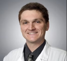
Richard Alan Rison is the Editor-in-Chief of Journal of Medical Case reports. He is also an associate neurology editor (editorial board) for BMC Neurology, and the former lead editor for case reports of BMC Research Notes (currently on the editorial board). His scholarly work focuses on medical case reporting, reporting standards, and editorial methodology. Dr. Rison participated in the development and dissemination of the CARE guidelines for clinical case reporting and has authored numerous publications addressing both neurological disorders and the role of case reports in advancing medical knowledge. Dr. Rison practices general neurology and served as the founding medical director of the PIH Health Hospital-Whittier Stroke Program and the PIH Health Hospital-Whittier Non-Invasive Vascular Laboratory, is a clinical assistant professor of neurology at the University of Southern California Keck School of Medicine and Los Angeles County Medical Center, and is a Fellow of the American Academy of Neurology, the American Neurological Association, and the American Association of Neuromuscular and Electrodiagnostic Medicine. Dr Rison is board-certified by the American Board of Psychiatry and Neurology in neurology and vascular neurology, and neurocritical care and neuroimaging by the United Council of Neurologic Subspecialties. He is also board-certified by the American Board of Electrodiagnostic Medicine in electrodiagnostic medicine. Dr. Rison is a former president of the Los Angeles Neurological Society.
Follow the Topic
-
Journal of Medical Case Reports
This journal will consider any original case report that expands the field of general medical knowledge, and original research relating to case reports.