Mind the gap on the way to eukaryotes
Published in Microbiology

Explore the Research

Spatial separation of ribosomes and DNA in Asgard archaeal cells - The ISME Journal
The ISME Journal - Spatial separation of ribosomes and DNA in Asgard archaeal cells
You sit in a dark room in front of a microscope and look for tiny and bright signals coming from your favourite bug. For hours, days, and maybe weeks…. Ok, congrats! You are lucky and patient enough to find a rare microbe! This makes your day. Or sometimes makes a paper….
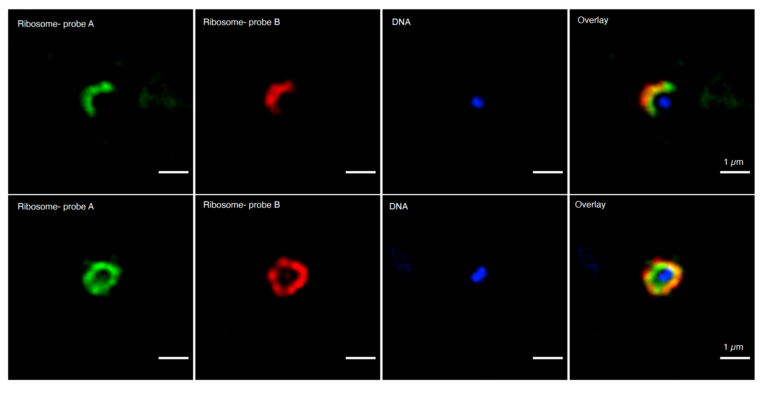
That pretty much sums up our recent study published in The ISME Journal. We imaged diverse Asgard archaeal cells in the marine environment for the first time. High-resolution microscopy revealed that DNA and ribosome signals separated in Asgard archaea cells, indicating a potential for cellular compartmentalization or membrane invagination.
How did the complex eukaryotic cells evolve?
The biology textbooks classify living organisms as prokaryotes or eukaryotes based on their cellular characteristics. Prokaryotic cells comprise bacteria and archaea, whose genetic material is not stored within a membrane-bound nucleus. On the other hand, eukaryotic cells are generally larger and more complex. They also contain a nucleus and a variety of membrane-enclosed compartments for localizing specific metabolic functions. The evolution of these complex cell types has long been a major open question in biology.
Archaea with eukaryotic tendencies
Recent analyses suggest that a merger between an Asgard archaeon and a bacterium gave rise to the first eukaryotic cell. Asgard archaea are the closest known extant prokaryotic relatives of eukaryotes. They also have several types of proteins previously considered specific for eukaryotes, suggesting a genetic potential for high cellular complexity. However, microscopic evidence to show the cellular structure of Asgard archaea in the environment is thus far lacking.
Hunting for Asgard archaea cells in marine sediments

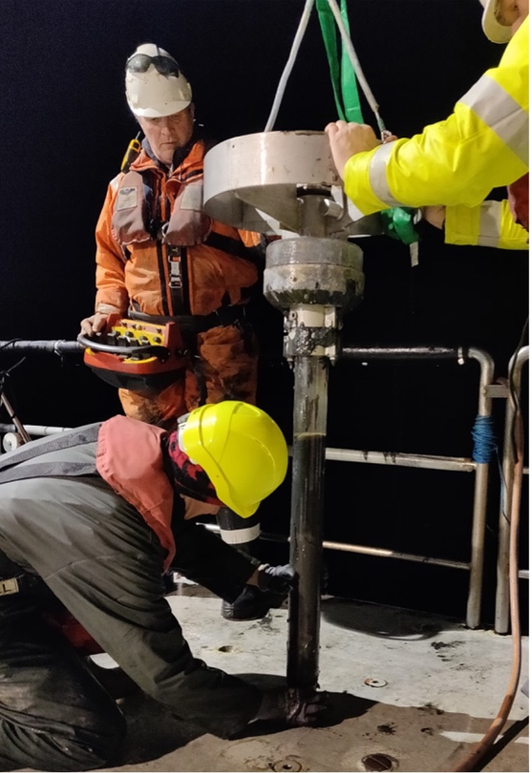
We organized several expeditions to sample marine sediments from Aarhus Bay, Denmark. We used a recently established primer-free rRNA sequencing method, which enabled us to obtain almost full-length 16S rRNA sequences of a diverse range of Asgard archaea populating the sediments. We then designed fluorescently labeled probes to specifically label Asgard archaea cells using catalyzed reporter deposition-fluorescence in situ hybridization (CARD-FISH). Finally, we imaged labeled cells using super-resolution microscopy.
The results were surprising: ribosome and DNA structures were spatially separated in Asgard archaeal cells. Honestly, it took some time to believe that these are “real” Asgard archaeal cells since DNA signals generally overlap partially or completely in prokaryotes. Comprehensive control experiments and reproducible signal patterns in several experiments revealed that these are obviously true positive Asgard archaeal cells.
A conspicuous gap between ribosomes and DNA

The results suggest that the genomic material is condensed and spatially distinct in a particular location in Asgard archaeal cells. Their genomes encode eukaryotic signature proteins involved in cytoskeleton formation and membrane remodelling. Therefore, this conspicuous gap between ribosomes and DNA in Asgard archaeal cells could hint a degree of cellular compartmentalization like eukaryotic cells.
Towards answering a puzzling question
Seeing is believing. Yet, these findings bring more questions to answer. For detailed imaging of cell interior, we will need to image Asgard archaeal cells using electron microscopy. This would provide insights into the emergence of subcellular complexity Asgard archaea and contribute to solving a major puzzle in the history of life.
Stay tuned! More to come soon…
Finally, I dedicate this paper to the memory of my beloved father Süreyya Avcı, who passed away during the preparation of this study. He always believed the dignity of science and thought me the essence of critical thinking. Şerefine Süreyya Hoca!

Avcı, B. et al. Spatial separation of ribosomes and DNA in Asgard archaeal cells. The ISME Journal 16, 606-610, doi:10.1038/s41396-021-01098-3 (2022).

Please sign in or register for FREE
If you are a registered user on Research Communities by Springer Nature, please sign in