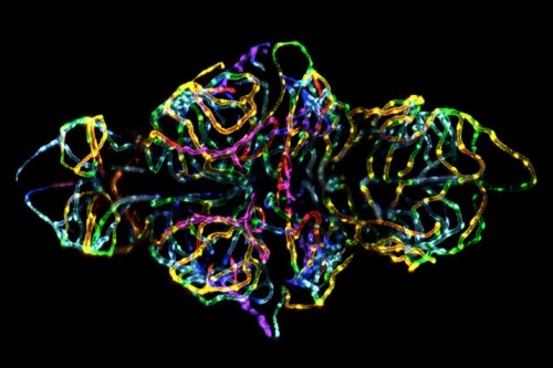
For the last few years Nature Methods has published the winning image of the Nikon Small World Photomicrography Competition on our cover. This year I was lucky enough to serve on the competition judging panel alongside three other judges chosen by the competition organizers. The competition was fierce but the image below was chosen as the 2012 winner.
The winning photomicrographers responsible for the image are microscopy specialist Jennifer Peters, a staff member of the Light Microscopy Core Facility at St. Jude Children’s Research Hospital in Memphis, Tennessee, and chemical biologist Michael Taylor. They created this image of the blood-brain barrier in a live transgenic zebrafish. The blood-brain barrier controls which substances come into contact with the vertebrate brain.
To view the other contest images and learn more about the competition please visit http://www.nikonsmallworld.com
Nature Methods talked briefly to the researchers about the science behind the beauty.
So what am I looking at?
MT: What you’re looking at is the blood-brain barrier of a live zebrafish larva at 6 days post-fertilization. The image of the brain vasculature is probably about 400 microns across.
JP: I always tell Mike that he has the most photogenic fish. It’s a 3D stack of images taken with a confocal microscope and collapsed in 2D. It changes from pink to red to yellow to green to blue as you go deeper into the brain. So the rainbow is pretty, but it’s also providing you with spatial information.
What do you hope to learn from these kinds of images?
MT: What we want to do with this is answer age-old questions in the field of brain-barrier biology. We want to answer “When does the blood-brain barrier develop, and what signaling pathways are involved in blood-brain barrier formation and maintenance?”
MT: We also want to be able to use it in drug screens. We’re interested in identifying compounds that modify the blood-brain barrier. The blood-brain barrier often keeps chemotherapeutics from getting into the brain, but it also breaks down in many neurodegenerative diseases. So if we can find small molecules that influence these processes, maybe we can come up with ways to treat diseases.
What did you have to do to get the image?
MT: We first made a transgenic blood-brain barrier reporter line in zebrafish to image this structure in a live animal. This transgenic line drives expression of the red fluorescent protein mCherry specifically in brain endothelial cells that make up the blood-brain barrier.
JP: In order to make the image look the way it does took some trial and error and some finessing. Imaging these live fish can be a challenge, since we have to keep the fish immobile and alive. We can also image the fish for up to 30 hours to examine development in real time.
These fish really are an ideal sample for microscopy and for the types of screens that Mike was talking about. You can fill up 96-well plates with fish embryos and visually screen them.
How did you get interested in science?
JP: [I’ve] always liked science ever since I was a kid. My father and I used to do chemistry experiments in the kitchen. I like instrumentation and like to take things apart and put them back together. That combined with the visual impact brought me to these kinds of studies.
MT: I had never considered science as a career. I was a business major and took chemistry to fulfill a requirement. At UC Davis, I became a biochemistry major and started working in a research lab to earn some spare money, and became interested in pursuing scientific research for my career. My boss encouraged me to go to graduate school, which is when I began working with zebrafish.


Please sign in or register for FREE
If you are a registered user on Research Communities by Springer Nature, please sign in