Researcher Riddle: Image manipulation
Published in Healthcare & Nursing, Astronomy, and Social Sciences

Many researchers include images in their manuscript – these can be images that are themselves the data or results of a study – such as western blots or images of mice tumours - or images such as graphs or charts that serve as visual representations of data. Images can be used to highlight key findings, emphasise and support arguments, and enhance reader comprehension. All in all, they are great tools for researchers to use in academic publishing and science communication. In today’s digital age, there is a wide variety of programs and methods at your disposal to alter and enhance your images for clarity and impact – but not all of them preserve the image’s integrity.
This week’s scenario quizzes you on acceptable methods of image modifications and below that, you can find a new resource on acceptable image modification.
Image modification policies consider various aspects of image production and editing, from questions regarding when and where images were taken, to acceptable and unacceptable methods of altering images.
Before we get into those, test your current knowledge of which image modification methods are compliant with Springer Nature policies – and which aren’t - below:
Which of the following examples are acceptable image modifications?
Select all that apply:
- A: A manuscript contains multiple Western blot images, two of which are almost identical to each other. One is missing some of the faint bands visible in the other, and in their place, the background is a lot lighter. This image shows signs of gel splicing, but this is not mentioned in the figure legend.
- B: Another manuscript also contains gel images, and notes in their figure legend that they have cropped these images for clarity. The authors have provided uncropped images in the supplementary information.
- C: A manuscript on a particular plant disease includes various images of crops. It mentions that these have been acquired through fluorescence imaging techniques and the authors state that the specific image acquisition settings and processing steps are available upon request.
- D: A manuscript contains a single figure comprised of three images. Each of the images has a time stamp in the right-hand corner. One of them seems to have been cropped as the time stamp is only half-visible – however, the month is still visible and this image is time stamped in the month of April, whilst the other two were taken in March.
-
E: A manuscript contains images of tumours grown in mice. The images have been adjusted in their entirety for brightness and contrast. No unaltered images were provided upon initial submission
- F: A manuscript is submitted containing western blot images. You notice that some of these images appear to be duplicated. . Upon closer inspection, it becomes clear that they are all supposed to represent β-actin loading controls. This reuse is not mentioned in the figure legend.

Image source: © BASILICOSTUDIO STOCK / stock.adobe.com
[Image description: A woman sits a table using a drawing tablet to interact with two monitors in front of her which show image editing software.]
The correct answers are B and E:
- Whilst cropping for clarity is allowed, this must be declared in the figure legend. It is important that authors are prepared to provide the original and uncropped images upon request – this also counts for all other acceptable modification methods, such as filtering for brightness and contrast. It is best practice to provide these images in the supplementary information from the first submission.
- As mentioned, adjustments such as brightening or enhancing contrast are allowed – however, they must be applied uniformly across the entire image. It is also important that these adjustments do not alter the visibility of the data presented, nor selectively emphasize certain features. Authors should be prepared to provide original, unaltered images if these are requested, or if this is not possible, duplicates or triplicates of the images.
As for the wrong answers:
- In option A, the undeclared image splicing would already be grounds for judging this modification as unacceptable. Furthermore, the situation hints at other breaches of image integrity policies as it sounds as though a part of the blot was either brightened so much as to render those faint bands invisible, or other digital image editing (e.g. a cloning tool) was used.
- In option C, the authors did not comply with the policy that all image acquisition tools, and software used should be documented, including version numbers. Image acquisition settings and processing steps should be fully documented in the Methods section.
- As for option D, the lack of visibility of the timestamp may be problematic, as images taken at different times should be clearly presented as such. For example, authors could use borders to clearly separate adjacent images and explain their relationship in the figure legend.
- In option F, there are some instances in which it is reasonable to reuse a control, however this should always be declared in the figure legend. A situation where it might be appropriate to reuse such control blots is when different proteins from the same gel are presented in separate figure panels. If reuse is undeclared, this should prompt further investigation.
Resources for image integrity concerns
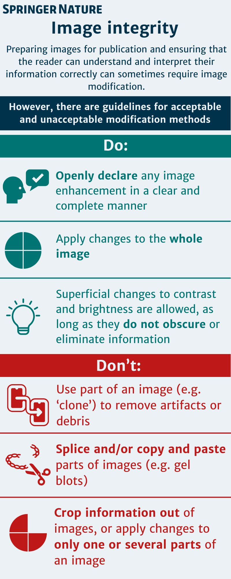
There are various resources at hand to support researchers with navigating our policies around image integrity, including a new infographic developed by the Springer Nature Research Integrity Group (SNRIG):
- Our handy new infographic, attached below, provides a bite-sized overview of acceptable image modifications. To save the image to your desktop, just right click and select ‘Save as image’. The infographic will also shortly be available on the policy page of the Springer Nature website – watch this space!
- This author tutorial page on Images provides a good oversight of the basic considerations to be made with various image types. This tutorial is linked to the broader Writing a Journal Manuscript course that is available to all researchers.

Please sign in or register for FREE
If you are a registered user on Research Communities by Springer Nature, please sign in