Respiratory tract commensal exacerbates allergic rhinitis
Published in Microbiology

Microbiological research in the last decade has discovered that the bacterial communities that live on epithelial surfaces of the human body – such as in the intestine or on the skin – have a considerable impact on human health. One site of bacterial colonization is the nose; and although there are by far not as many bacteria in the nose as in our gut, bacteria that colonize the nose have received much attention. This is because some of them, such as Staphylococcus aureus, which do not do any harm in the nose, can cause disease when they gain access to the bloodstream for example through nose picking and minor scratches in the skin. Bacteria in the nose thus have been attributed harmful or beneficial roles based predominantly on whether they can cause disease in remote locations or whether they inhibit or promote nasal colonization by such potentially harmful colonizers. In contrast, we do not know much about whether bacterial colonizers of the nose can directly impact diseases of the nose and upper respiratory system.
Nowadays, a common initial approach to test whether members of the colonizing microbiota may be associated with disease manifestations is to analyze the composition of the respective microbiota in healthy and diseased subjects at the respective colonization site. We were specifically interested in allergic rhinitis, or hay fever, a condition that plagues millions of people worldwide. Similar to other researchers before us, we analyzed the nasal microbiome in patients with allergic rhinitis and healthy controls but used a comparatively higher cohort to overcome contradictory results from those generally more limited earlier studies. To that end, Dr. Ping Miao, the first author, gathered samples together with Dr. Yiming Jiang at Shanghai Ren Ji Hospital/Shanghai Jiao Tong University School of Medicine for microbiome analysis (Figure 1).
.jpg) Figure 1. The figure shows Dr. Yimin Jiang (left) and Dr. Ping Miao (right) during patient consultation/sample acquisition at Shanghai Ren Ji Hospital’s Department of Otorhinolaryngology in 2018.
Figure 1. The figure shows Dr. Yimin Jiang (left) and Dr. Ping Miao (right) during patient consultation/sample acquisition at Shanghai Ren Ji Hospital’s Department of Otorhinolaryngology in 2018.
We were quite surprised to find not only a generally very different microbiome in patients with allergic rhinitis, but that one species stood out: Streptococcus salivarius, a species only very rarely associated with disease and even often promoted for its probiotic potential. As in any case of a finding revealing a very specific disease-associated dysbiosis, we were intrigued by the possibility that S. salivarius may be involved somehow in promoting or exacerbating symptoms of allergic rhinitis. Naturally, such dysbiosis results are only associative. It was entirely possible that S. salivarius was found more frequently in the noses of allergic rhinitis patients simply because it may “cope” better with the drastically different environment with, for example, strongly increased production of nasal mucus, rather than contributing causally to allergic rhinitis symptoms.
Therefore, to gain insight into a potential causal relationship between the presence S. salivarius and the severity of allergic rhinitis, we developed ex-vivo and in-vivo murine models to monitor the impact of S. salivarius on allergic rhinitis-related phenotypes. Much of this initial exploratory work was performed by Dr. Miao at the NIAID, in continuation of the long-standing collaboration of Dr. Min Li’s lab at Ren Ji Hospital and Dr. Otto’s lab at the NIAID. Having established protocols, Dr. Miao then returned to Shanghai to finish experiments.
The results from our laboratory experiments showed in fact that S. salivarius contributes to exacerbation of allergic rhinitis by inducing inflammation to an extent not seen with other bacteria (using S. epidermidis, another dominant nasal colonizer as control). As this was an intriguing and to some extent unexpected result, we wondered what distinguished S. salivarius from other bacteria in its capacity to cause inflammatory phenotypes associated with allergic rhinitis in the nose.
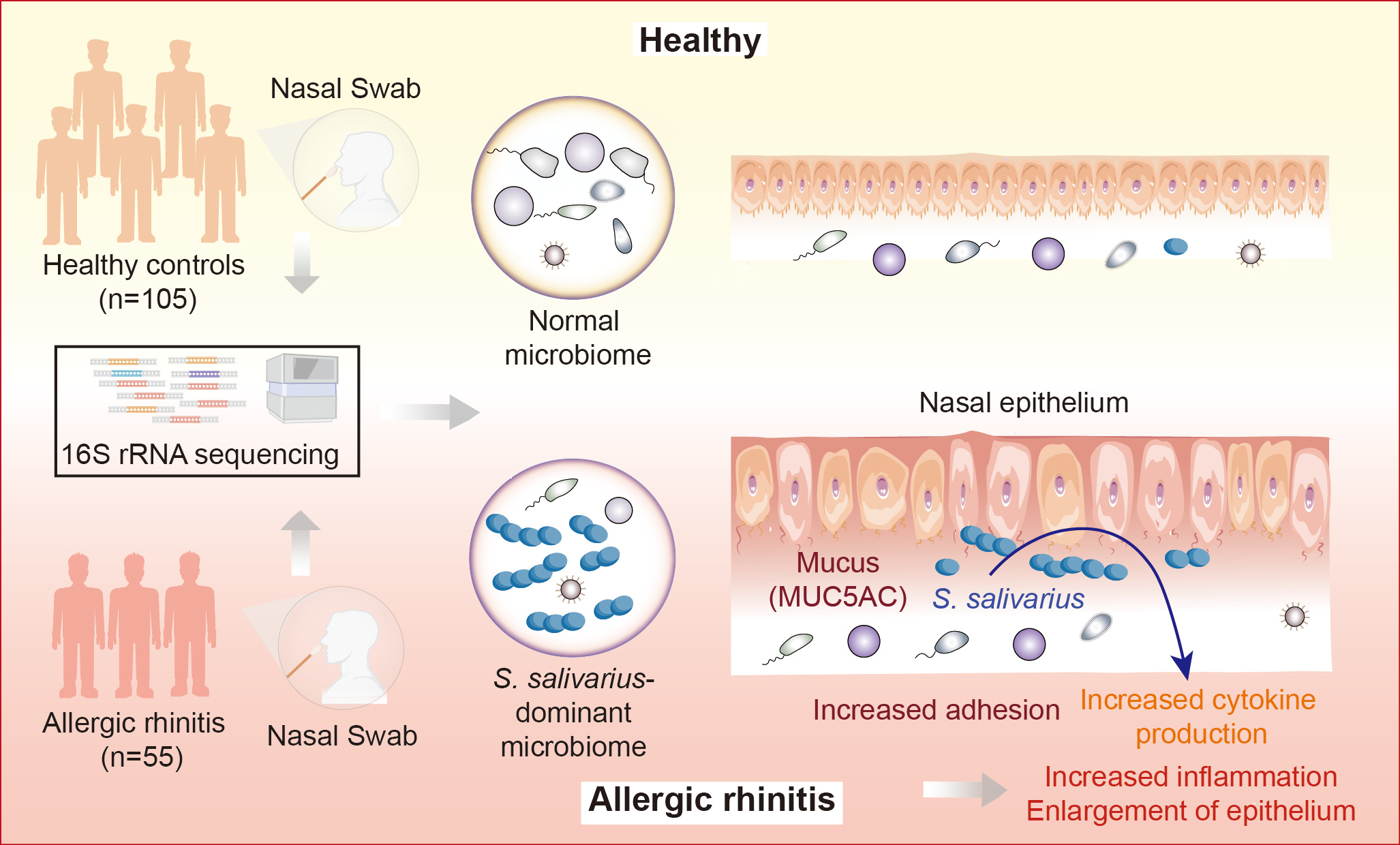
S. salivarius is not known to secrete exceptional pro-inflammatory molecules. Furthermore, in our in-vitro experiments only whole live bacteria but not culture filtrate had pro-inflammatory capacity. This led us to the hypothesis that exceptional adhesion to the epithelium rather than exceptional pro-inflammatory capacity is behind what we had observed. Indeed, S. salivarius showed strongly increased adhesion to allergen-exposed epithelial cells and lost allergic rhinitis-exacerbating capacity in mice that are devoid of the main nasal mucin Muc5ac, whose increased production is one of the hallmarks of allergic rhinitis. Thus, we believe that an exceptional capacity to adhere to Muc5Ac enables S. salivarius to remain in close contact with the nasal epithelium under conditions of allergic rhinitis, where then pro-inflammatory surface compounds (such as teichoic acids, lipoproteins etc.) induce inflammation. These molecules are not different from those in other bacteria, but as they require close contact of the bacteria with the epithelium, without the capacity to adhere to mucin – as exceptionally present in S. salivarius – they will have no pro-inflammatory effect (Figure 2).

Figure 2. The figure shows the setup of our microbiome analysis on the left and our model of how S. salivarius but no other bacteria exacerbate allergic rhinitis on the right. In allergic rhinitis, mucin (MUC5AC) is strongly overproduced, to which S. salivarius has exceptional capacity to adhere. This brings S. salivarius in close contact with the epithelium, triggering inflammation.
Certainly, this model is hypothetical to some extent and will need further experimental validation. This should include, for example, in-depth characterization of the role of the previously discovered S. salivarius Srp mucin adhesins in this context. We are also interested in further exploring the potentially beneficial role of S. epidermidis. Finally, the genus Subdoligranulum was also strongly associated with allergic rhinitis, but culturing of nasal isolates of this yet virtually unexplored genus has so far failed.
Follow the Topic
-
Nature Microbiology

An online-only monthly journal interested in all aspects of microorganisms, be it their evolution, physiology and cell biology; their interactions with each other, with a host or with an environment; or their societal significance.
Related Collections
With Collections, you can get published faster and increase your visibility.
The Clinical Microbiome
Publishing Model: Hybrid
Deadline: Mar 11, 2026


Please sign in or register for FREE
If you are a registered user on Research Communities by Springer Nature, please sign in