Scientific Maturation During a Study of Immature Flaviviruses
Published in Microbiology

You know how it goes. You’re at the beginning of your PhD, immersed in your main projects, when your supervisor comes to you with an idea: “Why don’t we try something on the side? Just a little side project.” Of course, you agree. You start working on it in your spare time, thinking it’ll be a nice addition. But then the first results come back… weird. You repeat the experiment – even weirder. You double-check your most basic protocols – still strange. At some point, you start to wonder: is something wrong with the methods? Or are we actually seeing something interesting?
This is how our story began – with tick-borne encephalitis virus (TBEV), a flavivirus from the same family as the more famous dengue, Zika, and West Nile viruses. These viruses share many features: they are small (around 50 nm in diameter), spherical, and enveloped, with a surface composed of two main structural proteins – envelope (E) and membrane (prM/M). These proteins form beautiful, icosahedrally symmetrical mosaics, giving flaviviruses a dangerous elegance. But while they look similar, some flaviviruses are spread by mosquitoes (like Zika and dengue), and others by ticks (like TBEV and its close relatives). And that raises a fascinating question: why are these viruses so vector-specific? Why can’t a mosquito-borne flavivirus infect ticks, or vice versa? Despite years of research, we still don’t fully understand why. It likely involves a combination of viral and vector-specific factors. But in our newly published Nature Communications study, we uncovered one unexpected difference between these two virus groups – and it all comes down to “immature” virus particles.
To explain what that means, here’s a quick tour through the flavivirus life cycle. Once a virus binds to a host cell receptor, it enters the cell via endocytosis. Inside the endosome, the drop in pH triggers a dramatic structural rearrangement of the viral surface proteins, exposing a fusion loop on the E protein that allows the virus to fuse with the endosomal membrane and release its genome into the cytoplasm – where replication begins.
Newly assembled virus particles are formed in the endoplasmic reticulum. At this stage, the particles are in a neutral pH environment and have a spiky appearance, thanks to the prM protein, which hasn’t yet been proteolytically cleaved. As the virions move through the Golgi, the acidic environment causes a reorganization of the surface proteins, exposing the cleavage site on prM for the host protease furin. Once furin cuts prM into pr and M, the pr segment remains attached, still shielding the fusion loop. Only after the virus is released into the neutral pH of the extracellular environment does the pr segment dissociate – “activating” the virus for its next infection. This is the textbook model. But as with many things in biology, reality is more complicated. Infected cells release not only fully mature particles, but also partially or fully immature ones. These immature particles form a sort of “structural quasispecies” whose behavior – and role in infection – is still poorly understood.
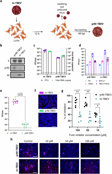
That’s where our story takes a twist. We set out to compare immature particles of mosquito-borne flaviviruses (MBFVs) and tick-borne flaviviruses (TBFVs), produced in LoVo cells – a cell line deficient in furin. What we found was surprising: immature TBFVs – including TBEV, Langat, and Louping ill viruses – remained fully infectious. Not just in cell culture, but also in live mice. In contrast, immature particles of MBFVs such as Zika, Usutu, and West Nile virus were almost completely non-infectious.
This was a striking result, especially since prM cleavage by furin was still required in both cases. So how could immature TBFVs be infectious?
.png)
The answer lies in the structure and dynamics of the E dimer – the paired arrangement of envelope proteins on the virus surface. Using mutagenesis, virological assays, structural modeling, and molecular dynamics simulations, we found that immature TBFVs have a unique ability to maintain a herringbone particle surface even at neutral pH, keeping the prM/E dimer interface stable and the furin cleavage site (FCS) exposed. That means furin can process the virus even after it has left the infected cell – on the surface of the next cell it tries to infect. MBFVs, on the other hand, tend to revert to the spiky, uncleavable form at neutral pH, effectively retracting their FCS and blocking further maturation unless they re-enter an acidic environment. This late-stage furin activation appears to be a special feature of TBFVs and may represent an evolutionary adaptation to their tick-borne transmission cycle. We identified key structural elements unique to TBFVs – including a longer stabilizing “fg loop” and specific interactions between E protomers – that help lock the E dimer in place and keep the FCS exposed. When we introduced mutations that made the E dimers more flexible, mimicking the MBFV-like behavior, we saw a loss of infectivity and a re-zippering of the prM region that retracted the FCS.
This led us to propose a new model: in TBFVs, immature particles circulate as “smooth - herringbone” rather than spiky virions, with prM loosely tethered to the surface. The FCS remains accessible, allowing furin on target cells to complete maturation during entry. This contrasts sharply with mosquito-borne viruses, where re-zippering and FCS hiding dominate unless acidic re-exposure occurs.
Why does this matter? First, it challenges the longstanding dogma that immature flaviviruses are uniformly non-infectious. Second, it reveals how subtle differences in structural dynamics can dramatically affect infectivity. And third, it hints at deeper evolutionary divergence between mosquito- and tick-borne flaviviruses – with implications for pathogenesis, immune evasion, and vector specificity.
Understanding these differences could also help guide future antiviral strategies, including vaccine development. If immature particles are more than just dead-end byproducts – if they can still infect under certain conditions – they may represent targets for therapeutic intervention. More broadly, this work reminds us that even “side projects” can lead to big insights – and that sometimes, when your results look strange, it’s not because you did something wrong… it’s because nature is showing you something new.
Follow the Topic
-
Nature Communications

An open access, multidisciplinary journal dedicated to publishing high-quality research in all areas of the biological, health, physical, chemical and Earth sciences.
Related Collections
With Collections, you can get published faster and increase your visibility.
Women's Health
Publishing Model: Hybrid
Deadline: Ongoing
Advances in neurodegenerative diseases
Publishing Model: Hybrid
Deadline: Mar 24, 2026


Please sign in or register for FREE
If you are a registered user on Research Communities by Springer Nature, please sign in