Severe diarrhea in newborns: Tiny tummy, big mystery
Published in Microbiology, General & Internal Medicine, and Immunology


The Viral Culprits
In the world of pigs, porcine enteric coronaviruses wreak havoc, causing severe gastroenteritis with infection rates soaring up to 100% in young piglets. These viruses, including Transmissible Gastroenteritis Virus (TGEV), Porcine Epidemic Diarrhea Virus (PEDV), and Porcine Deltacoronavirus (PDCoV), are a constant concern for the pig industry due to their high infection and mortality rates in young suckling piglets. Researchers have long puzzled over why these tiny tummies are so vulnerable. The answer might lie in the mucus—a gel-like barrier that lines the intestines and other hollow organs.
The Role of Mucus
Mucus is a major part of the innate immune system, covering the hollow organs of most mammals, including humans. It provides a barrier against normal bacteria present in the body and invading pathogens like viruses. This mucus is a gel that consists mainly of water, with large glycoproteins called mucins giving it its gel-like properties. It also contains other proteins, lipids, and electrolytes. Specialized cells in the inner lining of hollow tissues, called Goblet cells, are responsible for the formation of the mucus layer. This layer changes in terms of quality and quantity with age.
Age-Related Changes in Mucus
Studies have demonstrated a protective effect of mucus and mucins against disease-causing bacteria, viruses, and parasites. Mucus can either physically block pathogens by entrapment, inhibiting their access to target cells, or through chemical and immune entities present in the mucus. The changing parameters of these entities during infections can provide insights into their relation to disease pathogenesis. Researchers suspected that the severity of infection in young piglets could be related to these variations in mucosal components.
To investigate this, a study was conducted with four specific aims:
- To check the changes in the expression of important coronavirus receptors in three different ages of pig intestines.
- To investigate the changes occurring in intestinal morphology and mucus-producing cells of the intestines.
- To check the age-dependent blocking effect of intestinal mucus against TGEV.
- To see the age-related physical and chemical changes occurring in the pig intestinal mucus.
Changes in Intestinal Morphology and Mucus-Producing Cells
The study examined changes in intestinal morphology, the number of mucus-producing cells, and the expression of coronavirus receptors APN, DPP4, ACE2, and TMPRSS2 in pigs with aging. The expression pattern of these receptors was explored in four different regions of the intestines (duodenum, mid-jejunum, ileum, and colon) from three different ages of pigs (3 days, 3 weeks, 3 months). Immunofluorescence staining was used to visualize this pattern. The results showed that except for DPP4 in the jejunum, the expression of these receptors is highly variable and does not correlate with the age of the animal. Hence, the age-dependent susceptibility of animals to enteric coronaviruses is not dependent on the expression of these receptors.
This shifted the focus to changes in intestinal morphology and the number of mucus-producing cells. Using histochemical techniques, critical changes were observed in different regions of the intestines with aging. The main changes included an increase in the villus height-to-crypt depth ratio with aging, which affects nutrition absorption and mucus flow. The enterocytes, which are the main epithelial cells of the intestinal lining, increased in length with aging, which has significant implications for virus attachment and cell entry. Additionally, the number of mucus-producing cells increased with aging in all intestinal regions. These findings led to the conclusion that age-related changes occurring in intestinal mucus could help protect older animals against severe infections compared to newborn animals.
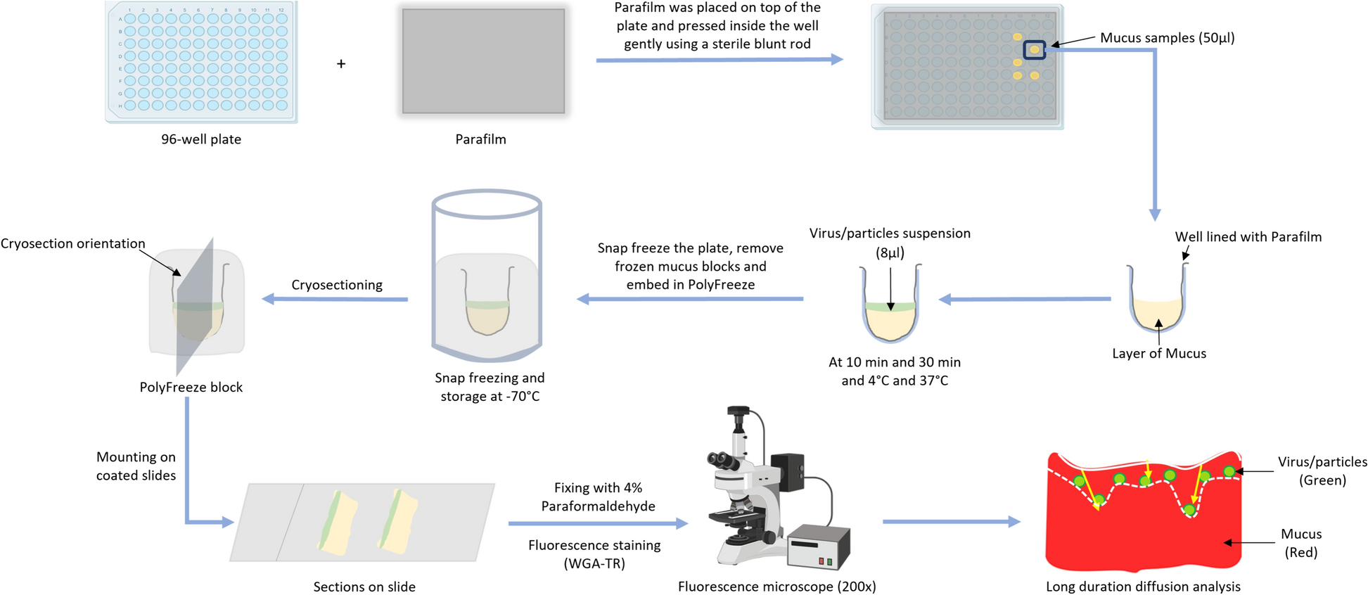
Protective Effect of Mucus Against Viral Infections
The age-dependent protective effect of porcine intestinal mucus against viral infections was explored in detail. The blocking effect of whole intestinal mucus extracted from 3-day and 3-week-old animals against TGEV was examined by tracking viral diffusion in the mucus and comparing the results with similar-sized, differently charged nanoparticles. The study showed that TGEV and control nanoparticles diffused freely in 3-day mucus while being hindered by 3-week mucus. Additionally, the protective effect of mucus against TGEV infection of susceptible ST cells was checked in the presence/absence of mucus. The results indicated that 3-week mucus has a significant TGEV-blocking activity compared to 3-day mucus, demonstrating the protective effect of intestinal mucus against viral infection and that the protective activity increases with age.
Physiochemical Properties of Mucus
The physiochemical properties of the mucus samples were also investigated. The flow pattern of mucus was measured in terms of viscosity using its rheological properties. The 3-week mucus exhibited more viscosity and less shear thinning compared to the 3-day mucus. Furthermore, mucus forms a meshwork of interlinked mucins that create variable-sized pores. Atomic force microscopy (AFM) was used to assess the topography of these surfaces. The mean pore size for 3-day mucus was higher than that of 3-week mucus, with more than 80% of the pores in 3-day mucus being larger than the average diameter of enteric coronaviruses compared to only 50% in 3-week mucus. Additionally, the protein profile of the mucus samples was assessed using mass spectrometry. Higher levels of MUC13 and APN were found in 3-day mucus, while increased MUC2 expression was observed in 3-week mucus. These parameters are known to support higher infection rates in young pigs. Thus, it was concluded that physiochemical differences between the intestinal mucus of 3-day-old and 3-week-old pigs show key differences that could explain the decrease in viral susceptibility of older pigs.
Conclusion
In conclusion, while the immune system and genetics play their roles, the often-overlooked mucus might just be the unsung hero—or villain—in this tiny tummy mystery. Understanding these changes could pave the way for new treatments and preventive measures, ensuring healthier beginnings for both piglets and human infants. The study highlights the importance of considering age-related changes in mucus composition and structure when studying infectious diseases, virus tissue tropism, and developing therapeutics.
Follow the Topic
-
Veterinary Research
This is an open access journal that publishes high-quality and novel research and review articles focusing on all aspects of infectious diseases and host-pathogen interactions in animals.

Related Collections
With Collections, you can get published faster and increase your visibility.
Addressing research gaps in foot-and-mouth disease: Insights from the Global Foot-and-Mouth Disease Research Alliance
Foot-and-mouth disease (FMD) is a highly contagious viral disease of ruminants, posing a persistent challenge to global animal health. Despite significant investments in control measures and vaccination, many countries continue to report frequent outbreaks, highlighting the need for improved strategies and innovative tools to combat the disease effectively.
FMD remains endemic in over 100 countries spanning Africa, Asia, West Eurasia, the Middle East, and South America. The ongoing emergence and re-emergence of viral strains, coupled with their transmission across borders, further complicates control efforts. According to the World Organisation for Animal Health (WOAH), among its 182 member countries, 68 are officially recognised as FMD-free (67 without vaccination and 1 with vaccination), while 13 countries have FMD-free zones with or without vaccination. However, 98 countries lack official FMD-free status, underscoring uneven progress toward global control.
Recognising the critical need to address gaps in knowledge, the Global Foot-and-Mouth Disease Research Alliance (GFRA) organised a workshop to conduct a comprehensive analysis of research gaps related to FMD control. This effort brought together international experts to evaluate the current state of knowledge and identify priorities for advancing countermeasures. The focus was on improving outbreak mitigation in FMD-free areas and supporting eradication efforts in endemic regions, addressing topics as pathogenesis, epidemiology, immunology, vaccines, antivirals and diagnostics.
Submissions to this Special Collection in Veterinary Research should address topics identified as research gaps in the Research Gap Analysis conducted by the GFRA in 2022.
Contributions are welcome in the form of full articles, short communications, or short reviews (limited to specific topics; see note below). These articles provide updates on key advances in FMD biology, pathogenesis, immune responses, and disease control strategies over the past five years. These reviews aim to guide scientists and policymakers in identifying future research priorities to enhance FMD management and alleviate its economic burden on livestock farmers worldwide.
By addressing these research gaps, the GFRA and its collaborators contribute to global efforts aimed at developing innovative tools and strategies to control and ultimately eradicate foot-and-mouth disease.
Note: Please contact the associate editors for approval of the review topic prior to submission.
This Collection supports and amplifies research related to SDG 2, Zero Hunger, SDG 3, Good Health and Well-Being, and SDG 15, Life on Land.
All submissions in this collection undergo the journal’s standard peer review process. Similarly, all manuscripts authored by a Guest Editor(s) will be handled by the Editor-in-Chief. As an open access publication, this journal levies an article processing fee (details here). We recognize that many key stakeholders may not have access to such resources and are committed to supporting participation in this issue wherever resources are a barrier. For more information about what support may be available, please visit OA funding and support, or email OAfundingpolicy@springernature.com or the Editor-in-Chief.
Publishing Model: Open Access
Deadline: Aug 31, 2026
Co-infections responsible for cattle respiratory diseases
Respiratory diseases are major causes of mortality, morbidity, and economic losses in cattle. Different pathogens, notably viruses and bacteria, are responsible for lower respiratory tract infections in bovines. By weakening their host, these pathogens facilitate opportunistic co-infections by other pathogens. In addition to the direct impact on animal health, these co-infections are major causes of the excessive use of antibiotics in farms. These treatments alter commensal flora of animals, which are known to participate in the biological equilibrium and immune status of organisms, thus increasing the risks of developing other illnesses. Furthermore, widespread and improper use of antibiotics in agriculture results in their release in the environment which favours the emergence of resistant bacteria and constitutes a serious threat for ecosystems and human health. These pressing concerns warrant a discussion concerning policies for antibiotics use in farms and livestock management practices to prevent such co-infections.
The objective of this series is to provide a comprehensive coverage of up-to-date research related to respiratory co-infections affecting cattle, in line with their impact on animal and human health, together with the preventive and curative measures that can be developed to control their impact. The series will focus on the most prevalent pathogens responsible for cattle disease, such as bovine respiratory syncytial virus, influenza D virus, bovine tuberculosis, and associated co-infections.
Veterinary Research invites you to submit articles on the topic of co-infections. To submit your manuscript, please use our online submission system, and indicate in your cover letter that you would like the manuscript to be considered for this article collection.
For more information about the journal and how to submit your article to Veterinary Research, please see our submission guidelines. Any accepted articles will appear together on this collection page, as and when the publications are ready.
To read our related collections on co-infections in animals, please see here.
Publishing Model: Open Access
Deadline: Ongoing


Please sign in or register for FREE
If you are a registered user on Research Communities by Springer Nature, please sign in