SproutAngio: An Open‑source Bioimage Informatics Tool for Quantitative Analysis of Blood Vessels
Published in Protocols & Methods
Changes in blood vessel morphology and endothelial cells are important to understand when developing a new biomedical application to treat a disease or when analyzing factors affecting neovessel formation during development or in disease. The new SproutAngio tool, developed in the University of Eastern Finland by Assoc. Prof. Johanna Laakkonen’s research group (@GroupLaakkonen), in collaboration with Profs. Jussi Tohka, Peetra Magnusson and Seppo Ylä-Herttuala, allows a detailed analysis of blood vessels from bioimages. In addition to improving the accuracy of several established image analysis methods, it provides new ways for automated and accurate analysis of endothelial lumen space and sprout morphology, a previously time-consuming part of the analysis.
The Solution to Automated Analysis of Endothelial Lumen Space and Sprout Morphology
New blood vessels are formed by angiogenesis, from pre-existing ones, or by vasculogenesis during development. SproutAngio is developed for analyzing bioimage data aiming to detect in detail vessel or endothelial sprouting in these processes. In our work, we combined existing sprout morphology analysis methods to our pipeline, but also introduced novel approaches including automated analysis of lumen space, while ensuring an easy use of the tool for researchers without prior programming experience. We prepared a detailed manual for the tool starting from the beginning with the installation of Python. By following the step-by-step manual, users can gradually learn how to modify several different parts of the analysis pipeline. The tool is open source, allowing for further improvements and customizations , while also providing researchers across the globe access to an easy, free-of-charge and automated image analysis tool.
 modelling angiogenesis in a cell culture, and vessels in mouse retina (right, scale bar: 200 µm) used for the development of the analysis tool are shown.")
SproutAngio is developed for analyzing endothelial and vessel structures. Example images from fibrin bead assay (left, scale bar: 100 µm) modelling angiogenesis in a cell culture, and vessels in mouse retina (right, scale bar: 200 µm) used for the development of the analysis tool are shown.
Comparison with Current Methods
Among similar image analysis tools, SproutAngio stands out for its 3D segmentation and automatic workflow of image processing, saving researchers valuable time. Users can easily check the resulting images to ensure the accuracy of segmentation and measurements before doing a batch analysis, i.e., automatically analyzing multiple images at a time. Moreover, SproutAngio’s Python-based approach offers unique advantages such as integrating machine learning-based bioimage analysis models, previously trained by other researchers, into the pipeline.
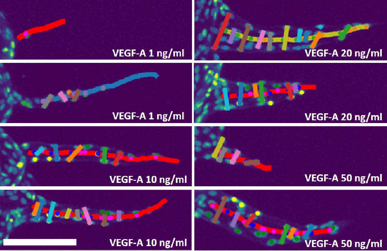
One of SproutAngio’s major advantages is providing two novel methods for the detection of lumen space, which removes the need for tedious and expensive immunolabeling protocols for detecting empty lumen space in vessels. Instead, SproutAngio measures lumen width from segmented images by analyzing the sprout width in various segments along the endothelial sprout or by measuring the paired nuclei distance using the nuclei data in the sprout.
Our Focus: FBA and Retina Datasets
In order to examine sprouting angiogenesis, we began by creating a carefully controlled dataset. We utilized an in vitro assay called the fibrin bead assay (FBA), which involved placing primary human umbilical vein endothelial cells (HUVECs) on collagen-coated cytodex beads and embedding them in fibrin gel, while fibroblasts were added on top. To evaluate our image analysis methods, we produced five groups with varying amounts of vascular endothelial growth factor A (VEGF-A) in the cell growth medium (ranging from 0 to 50 ng/ml) and used confocal microscopy to generate bioimage data. Using SproutAngio, we were able to perform a 3D segmentation and analyze the lumen space in novel ways, including regional width analyses and paired nuclei distance analyses.
Next, we tested the SproutAngio tool on tissue samples from a mouse retina model for a genetic disease, cerebral cavernous malformation (Ccm). We used Cdh5(PAC)-Cre-ERT2/Ccm3f/f mice (Ccm3iECKO), which had endothelial-specific and tamoxifen-inducible loss of function of the Pdcd10(Ccm3) gene. Our analysis focused on the branches close to the migration front, as vein enlargement and hypersprouting near this area were observed in previous studies on the Ccm retina model.
In vitro fibrin bead assay bioimage dataset used in the article is publicly available and can be downloaded for further method development and image analysis studies.

Overview of the datasets used for image analysis by the SproutAngio tool.
SproutAngio Improves Automated Analysis of Vascular Morphology
SproutAngio, our newly developed open-source tool, offers an excellent solution to the automated analysis of endothelial lumen space and sprout morphology. Compared to existing tools, it provides a more sophisticated and reliable analysis. SproutAngio's approach enhances the precision of automated analysis, making it easier to quantify the endothelial lumen space and to deduce technical quantitative analysis of vascular sprouts. We have tested and validated the tool through confocal microscopy datasets from in vitro and in vivo experiments showing that our novel methods introduced here are useful for studying vascular morphology. As automatized imaging is done more and more with histological scanners that then produce a large number of high-resolution bioimages, it is crucial to have proper automated image analysis tools to support the data processing. Our SproutAngio's approach provides an automated and reliable tool for the quantitation of angiogenic sprouts and vessel structures. It is open-source, accessible, and approachable to all researchers without programming experience! Go check it out: https://www.nature.com/articles/s41598-023-33090-6
Research interests: Cell-cell crosstalk in disease, angiocrine factors, mechanotransduction, vascular anomalies, aortic aneurysms, gene therapy
Follow the Topic
-
Scientific Reports
An open access journal publishing original research from across all areas of the natural sciences, psychology, medicine and engineering.

Related Collections
With Collections, you can get published faster and increase your visibility.
Health Policy and Systems Research
Publishing Model: Hybrid
Deadline: Jul 14, 2026
Healthy Aging
Publishing Model: Open Access
Deadline: Dec 31, 2026


Please sign in or register for FREE
If you are a registered user on Research Communities by Springer Nature, please sign in