Unlocking the Power of Deep Learning for Multi-Omic Molecular Profiling: Insights from a Pan-Cancer Study
Published in Cancer, Computational Sciences, and General & Internal Medicine
Snapshot
Cancer is a complex disease characterised by molecular alterations that can influence its progression and response to treatment. Molecular profiling tests are crucial for identifying these alterations and guiding treatment decisions. However, these tests are often time-consuming and expensive.
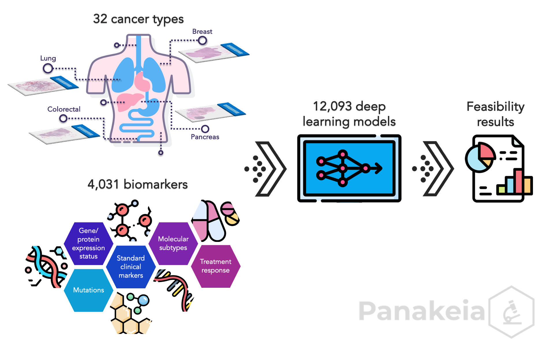
In our latest article published in Nature Communications Medicine, we present findings from a comprehensive pan-cancer study aimed at harnessing the potential of deep learning (DL) for molecular profiling through analysis of routine images of hematoxylin and eosin (H&E)-stained tissue samples. This in-depth investigation evaluated DL's capabilities to establish the status of over 4,000 multi-omic biomarkers across 32 cancer types. We trained and validated more than 12,000 DL models, covering a wide range of biomarkers across the molecular spectrum, such as mutations, gene and protein expressions, standard clinical markers, molecular subtypes, and treatment response.

Our key finding is that molecular profiling with image-based histomorphological features is generally feasible for most of the investigated biomarkers and across different cancer types. This is particularly significant because it showcases that DL can extract meaningful information from H&E-stained images that are correlated with molecular alterations. Another important finding is that the performance of DL models appears to be largely independent of tumour purity, sample size, and rarity of a biomarker.
The study also highlights the potential of DL to establish the status of biomarkers that are not routinely tested in clinical practice. For example, we found that some biomarkers that are associated with response to specific targeted therapies may be determined, which could enable personalised treatment decisions, once such approaches have been validated on larger cohorts.
Overall, the findings of our study demonstrate the promise of DL for establishing the status of multi-omic biomarkers directly from routine images of H&E-stained tissue samples. Clinical adoption of DL-driven approaches has the potential to accelerate diagnosis, guide treatment decisions, and improve outcomes for cancer patients.
Zoom-out
What is molecular profiling?
Molecular profiling is a process of identifying changes in certain molecules, such as genes, proteins, or metabolites, in a biological sample. This information can be used to understand the molecular basis of a tumour, identify potential therapeutic targets, and develop personalised treatments for patients. Molecular profiling is typically conducted in a laboratory and requires specialised, quality-assured work with dedicated equipment and expensive reagents.
What is deep learning?
Deep learning (DL) is a type of artificial intelligence that allows computers to learn from data without being explicitly programmed. DL may be used to train models to establish the status of biomarkers from digital images of H&E-stained histological slides. The models in our study were trained on a large dataset of images and corresponding biomarker data. Once trained, the models can be used for molecular profiling from new images.
What are the benefits of this approach?
There are several benefits to using DL for biomarker profiling from routine images of H&E-stained tissue samples. First of all, it is a fast and efficient method. The models can establish the status of biomarkers in a matter of seconds, significantly faster than traditional laboratory methods. Biomarker status can be established by analysis of existing H&E-stained slides, which eliminates the need for additional biopsies or tests. Finally, the cost of DL-based biomarker testing is significantly lower than the cost of traditional laboratory methods.
What are the implications of this research?
Showing the potential of DL-based biomarker profiling may be revolutionary for cancer diagnosis and treatment. By providing faster, more accessible, and more cost-effective molecular profiling tests, DL can help improve the lives of cancer patients worldwide.
About the authors
Panakeia Technologies provides comprehensive multi-omic molecular analysis of cancer patients significantly faster and cheaper than other alternative methods. We are building AI-technology to provide biomarker information directly from H&E-stained tissue images. Our solutions offer reliable results in minutes rather than several days or weeks, enhancing both clinical diagnostics and drug development.
Follow the Topic
-
Communications Medicine

A selective open access journal from Nature Portfolio publishing high-quality research, reviews and commentary across all clinical, translational, and public health research fields.
Related Collections
With Collections, you can get published faster and increase your visibility.
Reproductive Health
Publishing Model: Hybrid
Deadline: Mar 30, 2026
Healthy Aging
Publishing Model: Open Access
Deadline: Jun 01, 2026


Please sign in or register for FREE
If you are a registered user on Research Communities by Springer Nature, please sign in