When the “motor cerebellum” started to speak about social behavior
Published in Neuroscience and General & Internal Medicine
For a long time, the cerebellum has been considered a brain region primarily responsible for motor coordination. As neuroscientists, we were taught that if you want to study movement, you go to the cerebellum. If you want to study social behavior, you look elsewhere.
But over the past decade, that view has begun to change.
Accumulating evidence has suggested that the cerebellum is not just a “motor center,” but also plays roles in cognition, emotion, and even social behavior. This shift raised an important question for us: if the cerebellum contributes to social behavior, what are the underlying cellular and molecular mechanisms?
This question became the starting point of our study.
Two different models, one shared clue
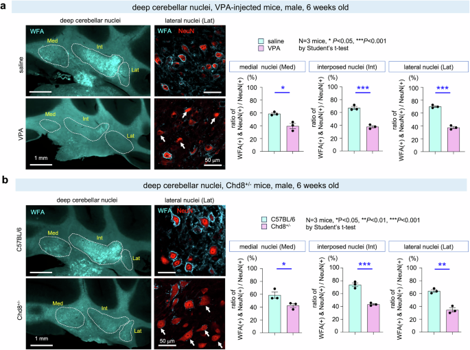
Autism spectrum disorder (ASD) is characterized by difficulties in social interaction and communication. It is highly heterogeneous, involving both genetic and environmental factors. To capture this complexity, we decided to work with two distinct mouse models:
- a prenatal valproic acid (VPA) exposure model
- a Chd8 mutant model
Although these models differ in origin, we reasoned that identifying shared changes across them could reveal core mechanisms underlying ASD-related phenotypes.
We began by surveying multiple brain regions. While expected changes were observed in some brain regions, what caught our attention was something unexpected.
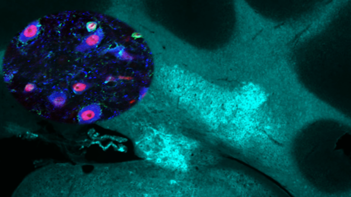
In both models, we found a reduction of perineuronal nets (PNNs) in neurons of the deep cerebellar nuclei.
This was surprising.
PNNs are extracellular matrix structures that wrap around neurons and help stabilize neuronal activity and synaptic function. Their role in the cerebellum, particularly in relation to social behavior, has remained largely unexplored.
This led us to ask:
Could changes in these extracellular structures in the cerebellum influence social behavior?
Breaking the net
To test this idea, we needed to move beyond correlation.
We used an enzyme to selectively degrade PNNs in the deep cerebellar nuclei. This allowed us to directly test their functional role.
The results were clear.
Mice with disrupted PNNs showed reduced interest in other mice and impaired social interaction. Importantly, these behavioral changes occurred without major motor deficits, indicating that the effect was specific to social behavior.
This was one of the key turning points of the project.
It suggested that PNNs in the cerebellum are not merely structural—they are functionally important for social behavior.
When c-Fos did not behave as expected
At this stage, we wanted to understand how neuronal activity in the cerebellar nuclei changes during social behavior.
A standard approach in neuroscience is to use immediate early genes (IEGs), such as c-Fos, as markers of neuronal activation. Naturally, we began with c-Fos.
However, this is where we encountered an unexpected challenge.
In cortical regions, c-Fos worked well and showed clear activity-dependent signals. In contrast, in the cerebellar nuclei, c-Fos signals were weak and did not reliably reflect behavioral activation. This made it difficult to interpret neuronal activity using conventional markers.
This discrepancy forced us to rethink our approach.
We then turned our attention to another activity-related marker: phosphorylation of CREB1, a transcription factor known to be activated by intracellular calcium signaling.
What we found was striking.
Unlike c-Fos, CREB1 phosphorylation showed a clear increase in cerebellar nuclei neurons after social interaction. Moreover, this signal was absent when PNNs were disrupted, indicating that neuronal activation itself was impaired.
This moment was particularly important for us.
It highlighted that different brain regions may require different readouts of activity, and that relying on standard markers alone can sometimes obscure key biological signals.
A circuit-level effect
With this new tool in hand, we examined how cerebellar activity influences the broader brain.
In control mice, social interaction induced robust activation in cerebellar nuclei neurons, and this activity propagated to downstream regions, including the midbrain and thalamus.
In contrast, in mice with disrupted PNNs, this activation was largely absent.
It was as if the cerebellum could no longer effectively “broadcast” its signals to the rest of the brain.
This finding suggested that a local structural change in the cerebellum can propagate through large-scale neural circuits, ultimately affecting behavior.
An unexpected molecular player
Further analysis revealed another unexpected finding.
Neurons lacking PNNs showed increased expression of ARNT2, a transcription factor involved in regulating neuronal activity. Notably, this increase was observed even under baseline conditions, suggesting a shift in the intrinsic state of these neurons.
We hypothesized that ARNT2 might mediate the effects of PNN loss.
When we suppressed ARNT2 expression, both neuronal activity and social behavior were restored.
This result connected extracellular structure (PNNs), intracellular regulation (ARNT2), and circuit-level function into a unified mechanism.
Rethinking the cerebellum in ASD
Traditionally, ASD research has focused on the cerebral cortex and synaptic dysfunction, while the cerebellum has been viewed mainly through the lens of motor control.
Our findings suggest a different perspective.
They indicate that the cerebellum, through its extracellular environment and output circuits, plays a direct role in regulating social behavior. They also highlight the importance of looking beyond neurons and synapses to include extracellular structures that shape neuronal function.
Looking ahead
Many questions remain.
Do similar mechanisms operate in the human brain?
How are PNNs regulated during development?
Can modulation of cerebellar circuits influence social behavior?
While we do not yet have all the answers, this study opens a new avenue for understanding the neural basis of social behavior and its alterations in ASD.
For us, this project began with a simple question—what is the cerebellum really doing? —and led to an unexpected connection between extracellular structures, neuronal activity, and behavior.
Sometimes, when familiar tools like c-Fos do not behave as expected, they guide us toward entirely new ways of seeing the brain.
Reference:
Follow the Topic
-
Translational Psychiatry
This journal focuses on papers that directly study psychiatric disorders and bring new discovery into clinical practice.

Related Collections
With Collections, you can get published faster and increase your visibility.
Moving towards mechanism, causality and novel therapeutic interventions in translational psychiatry: focus on the microbiome-gut-brain axis
Publishing Model: Open Access
Deadline: Nov 15, 2026

Please sign in or register for FREE
If you are a registered user on Research Communities by Springer Nature, please sign in