Assessment of guided lateral maxillary sinus lift procedure with simultaneous implant placement using stereolithographic surgical guide: a randomized controlled clinical study
Published in General & Internal Medicine
Abstract
Aim The aim of this study is to assess the efficacy of the stereolithographic surgical guide in reducing intraoperative and postoperative complication during lateral sinus lift operation.
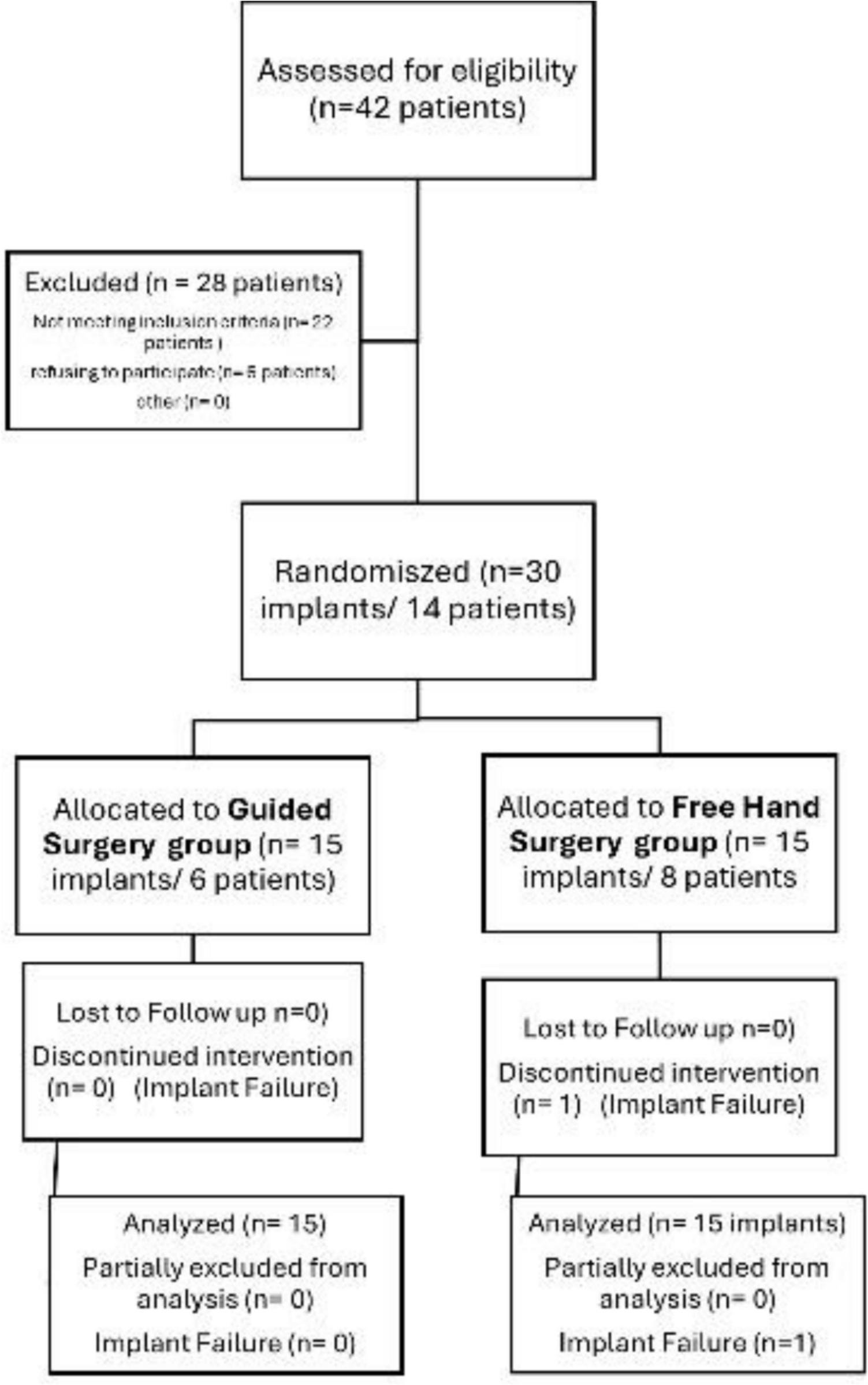
Materials and methods A parallel randomized controlled prospective clinical study was conducted on fourteen patients requiring thirty dental implants in the posterior maxilla and diagnosed with reduced vertical bone height. Lateral Maxillary Sinus Lift procedure with simultaneous implant placement was performed to all patients. Stereolithographic surgical guides for lateral window osteotomy and implant drilling and placement were used in the study group, while lateral osteotomy and implant drilling and placement was done freehand in the control group. A cone beam computed tomography was taken immediately and six months post-sinus lifting. Intraoperative and postoperative complications were assessed, pain and edema were assessed using visual analogue scale and vertical bone was assessed using fusion module of cone beam computed tomography.
Results
All dental implants demonstrated high survival rates with no statistically significant difference observed in intraoperative or postoperative complications. In terms of new vertical bone gain, both groups exhibited satisfactory and successful outcomes. Concerning pain, there was no statistically significant difference between the two groups except after two days, the study group showed statistically significantly lower pain score than the control group. While regarding the severity of edema, the study group showed statistically significantly higher prevalence of moderate and severe edema than control group which showed higher prevalence of mild edema.
Conclusion
According to the current study it has been concluded that there was no remarkable difference between the out-comes of both methods.
The study protocol and its consent form were approved by the ethical committee of Suez Canal University (No.432/2021); and registered retrospectively on 23 April 2024 on PACTR (PACTR20240875463218) (pactr.samrc.ac.za/TrialDisplay.
aspx?TrialID=30442).
Link to full article: https://link.springer.com/article/10.1007/s10006-025-01399-3
Follow the Topic
-
Oral and Maxillofacial Surgery
This is a peer-reviewed online journal that caters to the needs of professionals in oral and maxillofacial surgery.

Related Collections
With Collections, you can get published faster and increase your visibility.
Current State and Future Opportunities for 3D Printing/Additive Manufacturing in Oral and Maxillofacial Surgery: from Basic Science to Clinical Applications
Publishing Model: Hybrid
Deadline: Ongoing
Please sign in or register for FREE
If you are a registered user on Research Communities by Springer Nature, please sign in