Calcium Influx via LTCC Regulates Cardiac Regeneration
Published in Biomedical Research
The problem : the ever-increasing global burden of heart failure
Roughly 100.000 times a day the heart beats to pump blood through our body to disperse nutrients and oxygen to other organs and tissues, a ‘simple’ task yet critical for sustaining life. A disturbance in the force or efficiency with which the heart contracts has debilitating, widespread consequences. One of the main causes responsible for an impaired cardiac function is a myocardial infarction (heart attack), where blood flow to part of the heart muscle is blocked. In the absence of sufficient blood flow, cardiac tissue, including the cells chiefly responsible for heart contraction (cardiomyocytes), dies, leaving behind a path of destruction in the affected area. Given the importance of the heart in keeping us alive, surely there is an effective strategy to repair this damage and restore function, right? The short answer is not really. The affected area gets patched up by scar tissue produced by fibroblasts to prevent rupture of the heart muscle. However, this tissue does not actively contribute to heart contraction due to the absence of healthy cardiomyocytes. Why not make new cardiomyocytes you may ask? While the developing mammalian heart grows through the proliferation of the heart muscle cells, shortly after birth these same cardiomyocytes undergo a process of maturation and do no longer actively partake in the cell cycle nor can this process be naturally reactivated in response to cardiomyocyte death 1. Hence, the mammalian heart does not regenerate functional cardiac tissue. Over 60 million people worldwide live with heart failure 2, and while drugs are available to slow down disease progression, a cure that induces repopulation of the damaged tissue with functional heart muscle cells, thereby restoring the pump function of the heart, is yet to be developed.
The goal: understanding cardiac cell cycle regulation in response to changes in calcium flux
Fluxes of calcium in and out of the cell regulate the contraction of cardiomyocytes. But besides this, calcium is also an important signaling molecule and has been linked to the cardiac cell cycle 3. However, how this works exactly is still unknown. In our study, we used different in vitro and in vivo models to gain insights into the role of calcium in regulating the cell cycle in cardiomyocytes.
The results – a calcineurin-dependent route to cardiac proliferation
We started off with a drug screen targeting different phases of the cardiac calcium cycle. For these studies, we used human cardiac organoids, which are mini 3D models of the heart incorporating many important cardiac cell types (cardiomyocytes, fibroblasts, endothelial, and epicardial cells). Multiple aspects were investigated including 1) the initial influx into the cell via L-type calcium channels (LTCC), 2) the influx of calcium from the intracellular calcium store (sarcoplasmic reticulum) through ryanodine receptors, 3) the re-uptake of calcium into the sarcoplasmic reticulum via Sarcoendoplasmic Reticulum Calcium ATPase (SERCA), and 4) overall calcium by manipulating the calcium concentration in the culture media. Interestingly, we show that it is the initial influx of calcium that holds a grip on the cardiac cell cycle. These findings are consistent with the results obtained by Woo et al 3 and were confirmed in our study using primary mouse cardiomyocytes, so this was a robust and reproducible result.
Remarkably, cardiomyocytes express an endogenous inhibitor of LTCC called RRAD. Using single-cell RNA sequencing to analyze which genes are expressed in individual neonatal cardiomyocytes that are known to still have proliferative capacity, we found that RRAD was highly expressed in immature cardiomyocytes and cardiomyocytes that respond to a pro-proliferative cocktail. To confirm its role in cell cycle regulation, overexpression of RRAD resulted in increased cell cycle activity of cardiomyocytes (in vitro) and made these cardiomyocytes more responsive to pro-proliferative cocktails. These findings were further confirmed in adult human heart slices (ex vivo) and in mice (in vivo), supporting the robustness of this response across different species and levels of cardiac maturity.
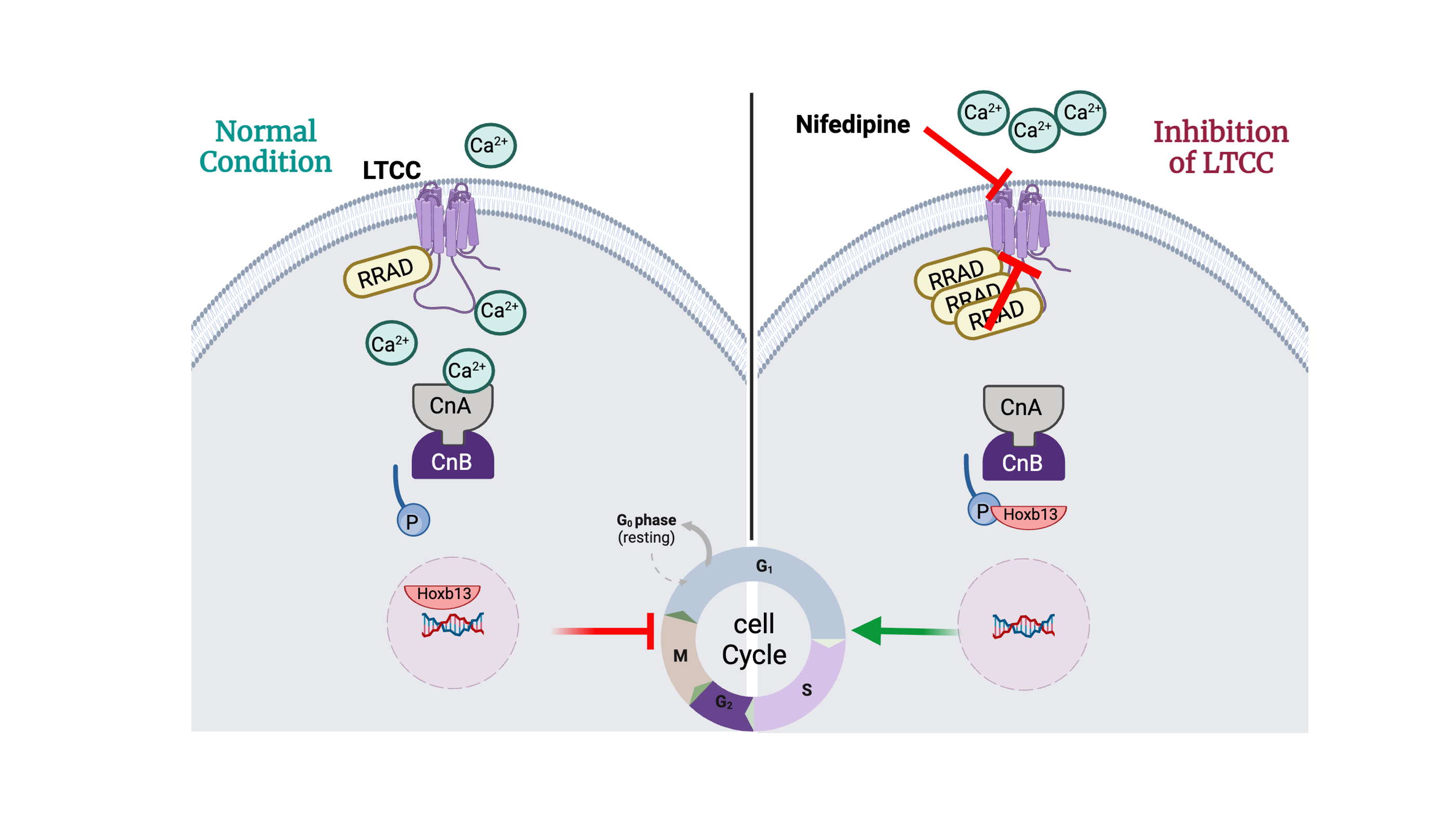
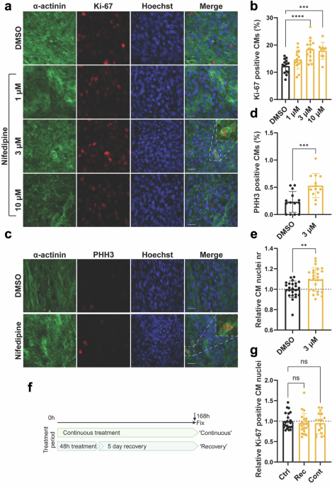
How does this change in calcium influx induce the cell cycle? Within cardiomyocytes, there are numerous proteins that sense and respond to changes in calcium concentration, thereby relaying information to the nucleus to regulate which genes get expressed. Calcineurin is a calcium-sensing protein that has been linked to the cardiac cell cycle previously 4,5, and in response to RRAD overexpression calcineurin activity was reduced. The activity of calcineurin transcriptional regulation is indicated by the expression of Rcan1, a protein that regulates calcineurin activity in a negative feedback loop6 which we found was decreased with RRAD overexpression. But how does calcineurin control the cell cycle? We found that a downstream target of calcineurin, HOXB13, which is directly involved in blocking cell cycle gene activity 5, was translocated out of the nucleus following RRAD overexpression. The same results were obtained when LTCC activity was inhibited using small molecule nifedipine. Combined, these data led us to the conclusion that calcium influx through LTCC regulates cardiac proliferation through calcineurin, which in turn regulates HOXB13 nuclear localization and cell cycle gene expression.
With the ultimate goal being to induce cardiac regeneration in the failing heart, we studied the impact of RRAD overexpression following an induced myocardial infarction in vivo in mice. We found that this alone was insufficient to repair the damage after myocardial infarction. But when RRAD overexpression was combined with the overexpression of two key cell cycle regulators (CCND and CDK4), scar size was reduced and contractile parameters improved, indicating that RRAD overexpression is a promising tool to enable repair of the heart post myocardial infarction
Future perspectives – identification of viable targets for regenerative therapeutics
Elucidating the why’s and the how’s of the endogenous regulation of the cardiac cell cycle is the first major hurdle that needs to be taken in the quest to a successful regenerative therapeutic. Our research shows how inhibition of LTCC using RRAD overexpression in combination with overexpression of two cell cycle genes could help repair the heart after a heart attack, which builds on previous research and solidifies cardiac calcium flux as a promising target to manipulate cardiac proliferation. The next challenge is to identify ways to safely manipulate the calcium influx or the downstream signaling events without interfering with cardiac function while still eliciting a cell cycle response.
References
1 Porrello, E. R. et al. Transient regenerative potential of the neonatal mouse heart. Science 331, 1078-1080 (2011). https://doi.org/10.1126/science.1200708
2 Groenewegen, A., Rutten, F. H., Mosterd, A. & Hoes, A. W. Epidemiology of heart failure. Eur J Heart Fail 22, 1342-1356 (2020). https://doi.org/10.1002/ejhf.1858
3 Woo, L. A. et al. High-content phenotypic assay for proliferation of human iPSC-derived cardiomyocytes identifies L-type calcium channels as targets. J Mol Cell Cardiol 127, 204-214 (2019). https://doi.org/10.1016/j.yjmcc.2018.12.015
4 Lam, N. T. et al. Targeting calcineurin induces cardiomyocyte proliferation in adult mice. Nat Cardiovasc Res 1, 679-688 (2022). https://doi.org/10.1038/s44161-022-00098-6
5 Nguyen, N. U. N. et al. A calcineurin-Hoxb13 axis regulates growth mode of mammalian cardiomyocytes. Nature 582, 271-276 (2020). https://doi.org/10.1038/s41586-020-2228-6
6 Yang, J. et al. Independent signals control expression of the calcineurin inhibitory proteins MCIP1 and MCIP2 in striated muscles. Circ Res 87, E61-68 (2000). https://doi.org/10.1161/01.res.87.12.e61
Follow the Topic
-
npj Regenerative Medicine
This journal is an open access, online-only, peer-reviewed journal dedicated to publishing high-quality research on ways to help the human body repair, replace and regenerate damaged tissues and organs.

Related Collections
With Collections, you can get published faster and increase your visibility.
Repairing and Regenerating the Vascular Code Across Tissues and Time
Publishing Model: Open Access
Deadline: Jul 18, 2026
Cellular and Genetic Tools in Regenerative Medicine
Publishing Model: Open Access
Deadline: Sep 11, 2026


Please sign in or register for FREE
If you are a registered user on Research Communities by Springer Nature, please sign in