Can AI-Supported Digital Microscopy Bridge the Diagnostic Gap in Primary Health Care?
Published in Healthcare & Nursing, Social Sciences, and Protocols & Methods


The World Health Organization (WHO) has highlighted how crucial it is to bring diagnostics closer to patients in primary health care. Yet many clinics in resource-constrained settings still lack access to basic laboratory testing. When accurate tests can be performed at the point of care, results are usually achieved faster, which improves treatment decisions and reduces the risk of diagnostic errors.
Manual microscopy remains a cornerstone of diagnostics in primary healthcare as it is affordable, versatile, and allows clinicians and laboratory staff to directly visualize pathogens and cellular changes in the sample. However, high-quality microscopy depends on skilled personnel and reliable infrastructure, both of which are limited in primary healthcare settings with constrained resources. This can lead to large differences in diagnostic quality and access between facilities and regions.
AI-supported digital microscopy is emerging as a powerful technology for laboratory diagnostics and may be particularly valuable in primary healthcare. By automating parts of the microscopy workflow and reducing reliance on on-site experts, it can help overcome limitations of traditional manual microscopy. This has the potential to improve access to diagnostic services where they are needed most, especially in expert-scarce settings such as low- and middle-income countries and sparsely populated areas in high-income countries.
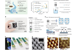
In the scoping review, the researchers identified articles investigating complete end to end AI-supported digital microscopy in primary health-care laboratories world-wide. End-to-end AI-based diagnostics here refers to a complete diagnostic workflow from collecting and digitizing samples to generating diagnostic results on a sample level. This was done through a systematic search of four scientific databases where articles were identified that reported results on diagnostic accuracy using digital microscopy, AI, and being performed in primary health care laboratories. A total of 3,403 articles were identified during the initial search, of which 22 (0.6%) were included after the screening process in the scoping review.
The results of the scoping review highlighted a broad applicability of AI-supported digital microscopy: “What stands out in our review is the breadth of applications where AI-supported digital microscopy shows promise, ranging from complete blood counts and malaria detection to stool parasites and cervical cancer screening” says Nina Linder, senior researcher from FIMM and professor at Uppsala University. “Many of the applications focus on conditions that place a disproportionate burden on vulnerable groups, for example cervical cancer screening among women and different parasitic infections among children” says Professor Linder. “If we can bring these AI-based tools into primary health care and point-of-care settings, we have a real opportunity to narrow diagnostic gaps for those who need it most,” says Professor Johan Lundin, senior co-author from FIMM and Karolinska Institutet.
Another main finding was the high diagnostic accuracy achieved with AI-supported digital microscopy. “In our review, AI-supported digital microscopy showcased diagnostic accuracy comparable to manual microscopy” says Dr Joar von Bahr, first author from FIMM and Karolinska Institutet. “Particularly encouraging was that AI-supported systems showed higher sensitivity than manual microscopy in six out of seven studies that made this comparison. This level of performance suggests that AI-supported digital microscopy may ultimately be used to enhance the diagnostic accuracy in primary healthcare” says von Bahr.
“Although AI-supported digital microscopy shows great promise, our review shows that the next step is to move beyond pilots and develop solutions that are scalable, transferable, and truly workable in routine practice. These systems ought to be cost-effective, and easy to implement across diverse laboratories,” says Professor Linder.
The study was carried out in collaboration between the Institute for Molecular Medicine Finland (FIMM) at the University of Helsinki, Karolinska Institutet, Uppsala University, and was supported by the Erling-Persson Foundation, Wallenberg Autonomous Systems and Software Program (WASP) funded by the Knut and Alice Wallenberg Foundation, the Swedish Research Council, and Finska läkarsällskapet, Liv och Hälsa, Sigrid Jusélius foundations in Finland.
-
I have a medical degree from Karolinska Institutet and am currently a PhD student at Karolinska Institutet and the University of Helsinki under the supervision of Professors Nina Linder and Johan Lundin.
-
The focus of my PhD is developing artificial intelligence supported image-based diagnostics for low-resource settings. Primarily for cervical cancer screening and soil-transmitted helminth diagnosis.
-
I am interested in the integration of medicine and technical advancements to improve diagnosis, especially with a focus on artificial intelligence.
- https://ki.se/en/people/joar-von-bahr#about-me

Please sign in or register for FREE
If you are a registered user on Research Communities by Springer Nature, please sign in