Detecting Heart Disease Smarter: Trade-offs in Accuracy, Speed, and Energy Use
Published in Computational Sciences


Cardiovascular diseases (CVDs) remain a major global health concern, contributing significantly to mortality and placing a heavy burden on healthcare systems. Early detection is key to improving patient outcomes, and electrocardiogram (ECG) signals have long been used as a reliable, non-invasive method for identifying cardiac abnormalities. However, interpreting these signals accurately requires expert knowledge and can be time-consuming, prompting the need for automated tools that streamline the diagnostic process.
In paper, "Machine Learning-Based Detection of Cardiovascular Disease Using ECG Signals: Performance vs. Complexity," Pham et al. explore how machine learning techniques can be applied to ECG analysis to improve both accuracy and efficiency. The study compares several approaches—Poincaré diagram-based image classification, 1D convolutional models, and XGBoost with engineered features—to determine which methods offer the best balance between performance and computational cost.
Understanding the Data
The researchers evaluated their models using two publicly available datasets from the PhysioNet/Computing in Cardiology Challenges: CinC 2017 and CinC 2020. These datasets contain ECG recordings from a wide range of patients and include labels for various types of cardiac conditions. CinC 2017 focuses specifically on arrhythmias, particularly atrial fibrillation (AF), while CinC 2020 includes a broader spectrum of cardiovascular diseases across multiple ECG leads.
These datasets are valuable not only for their size and diversity but also because they reflect real-world variability in ECG readings. The authors note that class imbalance is common in such datasets, with some conditions appearing far more frequently than others—a challenge that must be considered when evaluating model performance.
Exploring Different Modeling Approaches
Poincaré Diagrams
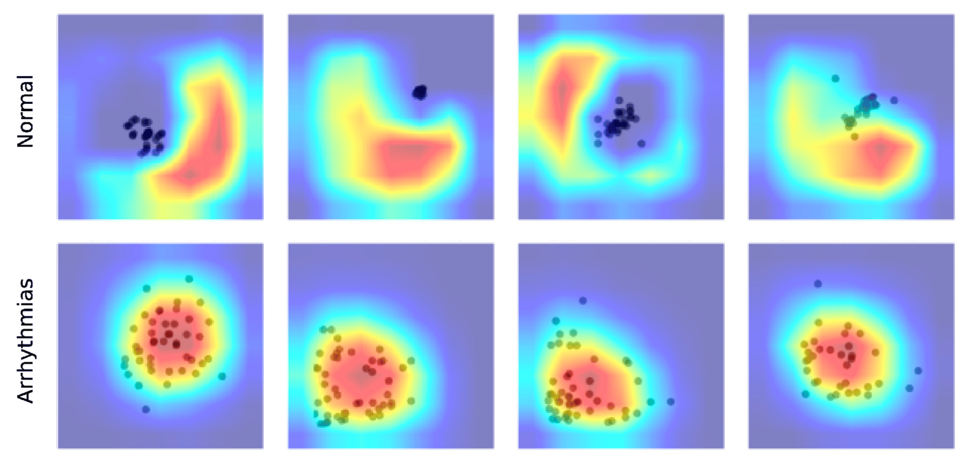
One approach examined was the use of Poincaré diagrams, which visualize heart rate variability by plotting consecutive R-R intervals from an ECG signal. These diagrams were then classified using deep learning models like ResNet50 and DenseNet121, typically used for image recognition tasks.
While this method showed promise in detecting AF, it struggled with other types of arrhythmia. This suggests that while heart rate variability is informative for certain conditions, it may not capture enough detail for broader disease classification. Still, the simplicity of generating Poincaré diagrams could make them useful in settings where advanced equipment is not available.
1D Convolutional Models
The most effective results came from applying 1D convolutional neural networks (CNNs) directly to raw ECG signals. Specifically, the 1D ResNet architecture outperformed existing solutions in both the CinC 2017 and CinC 2020 challenges, achieving F1 scores of 85% and 71%, respectively.
These models benefit from residual connections, which help maintain performance even as network depth increases. Importantly, the models also demonstrated strong specificity and consistent performance across different subsets of data, indicating robustness in real-world applications.
Additionally, these models consumed less power during inference compared to image-based approaches, making them suitable for deployment on wearable or portable devices where energy efficiency is crucial.
XGBoost with Engineered Features
An alternative approach involved extracting time-series features from ECG signals and training an XGBoost classifier. While this method performed reasonably well, especially on longer ECG recordings, its inference speed was notably slower due to the computational cost of feature extraction.
Feature importance analysis revealed that peak-related features, such as frequency components and statistical measures, played a significant role in prediction. However, the preprocessing overhead made this method less favorable for real-time monitoring scenarios.
Efficiency and Environmental Considerations
Beyond predictive accuracy, the study also assessed each model's computational efficiency, including inference time, power consumption, and carbon emissions. Not surprisingly, image-based models trained on Poincaré diagrams were among the most resource-intensive, while 1D convolutional models stood out for their low energy use.
This focus on environmental impact is timely, as concerns about the sustainability of AI grow. By quantifying CO₂ emissions associated with model training and inference, the authors provide insight into how machine learning can be deployed responsibly in clinical settings.
Interpreting Model Decisions
Understanding why a model makes a particular prediction is essential in medical contexts. To this end, the authors used GradCAM to visualize which parts of the ECG signal influenced the model’s decisions.
For the 1D ResNet model, attention was focused around the QRS complex, along with the P-wave and T-wave—regions known to be clinically relevant for diagnosing arrhythmias. This alignment with human expertise adds confidence in the model's reliability.
Similarly, DenseNet121 applied to Poincaré diagrams appeared to rely on patterns consistent with established understanding of heart rate variability, further supporting the interpretability of the results.
Practical Implications
The findings suggest that 1D convolutional models, particularly those based on ResNet architecture, offer a compelling combination of performance, efficiency, and interpretability for ECG classification. Their ability to process raw signals without extensive preprocessing makes them well-suited for integration into wearable health monitoring systems.
Meanwhile, the Poincaré diagram approach, though less accurate overall, remains a viable option for environments with limited resources. Further refinement of this method could expand its utility in point-of-care diagnostics.
The XGBoost model, while interpretable and effective on long-term data, faces practical limitations due to its computational demands. Optimizing feature selection or integrating lightweight preprocessing pipelines could enhance its viability for real-time use.
Looking Ahead
As machine learning continues to mature in the healthcare space, balancing performance with computational and environmental costs becomes increasingly important. Pham et al.'s work contributes to this conversation by offering a comprehensive comparison of modeling strategies for ECG classification.
Future research could explore hybrid approaches that combine the strengths of different models—for instance, integrating CNNs with attention mechanisms or developing lightweight versions of transformer-based architectures for ECG processing.
Moreover, expanding interpretability tools beyond GradCAM and feature importance could foster greater trust in AI-assisted diagnosis. As models become more integrated into clinical workflows, ensuring transparency will be key to gaining acceptance among healthcare providers.
Conclusion
Pham et al. present a thorough evaluation of machine learning techniques for ECG-based cardiovascular disease detection. Their results underscore the value of 1D convolutional models in delivering high performance with minimal resource use. By addressing both technical and practical considerations—including model complexity, energy efficiency, and interpretability—the study offers a roadmap for deploying machine learning in real-world cardiac diagnostics.
As ECG monitoring becomes more accessible through wearable technology, these findings pave the way for smarter, more scalable tools that support clinicians and improve patient care.
PhD in Computational Physics, Head of R&D at Sber AI, Scientific consultant at AIRI. Alma mater: MIPT 2019, ESPCI 2014, NSU 2011.


Please sign in or register for FREE
If you are a registered user on Research Communities by Springer Nature, please sign in