Detecting p53 Aggregates for Cancer Diagnosis
Published in Protocols & Methods and General & Internal Medicine

From Molecular Mystery to Clinical Potential
Proteins are the workhorses of the cell. If we imagine a cell as a tiny factory, proteins are its highly skilled workers, each performing dedicated roles to keep the system running smoothly. But what happens when some of these workers start clumping together and refuse to do their jobs?
This is the essence of protein aggregation, a process well-known in neurodegenerative diseases (NDs), such as Alzheimer’s and Parkinson’s. In these conditions, misfolded proteins lose their original functions and form aggregates that can disrupt cellular processes. Beyond NDs, emerging evidence suggests that protein aggregation also plays a role in cancer, particularly involving the well-known tumour suppressor p53.
p53 Aggregation and Cancer
Nicknamed “the guardian of the genome”, p53 is one of the most studied proteins in cancer biology, playing a key role in responding to cellular stress and preventing tumour growth. However, both wild-type (WT) and mutant forms of p53 can aggregate under stress, potentially losing their tumour-suppressing function and even exhibiting tumour-promoting properties.
In our previous collaboration with Prof. Kevin Brindle, we demonstrated that full-length WT p53 and its R248Q mutant can aggregate and disrupt lipid membranes, offering a novel mechanism for tumour development that does not rely solely on p53 mutation. This prompted an intriguing question: can p53 aggregates serve as a diagnostic marker for cancer?
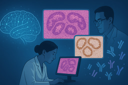
Development of an Immunoassay
We began to design an immunoassay capable of detecting p53 aggregates in liquid biopsy samples, such as blood plasma. To achieve high sensitivity and specificity, we first compared two single-molecule techniques: Single-Molecule Pull-down (SiMPull), a fluorescence imaging-based method, and Single-Molecule Array (SiMoA), a commercially available platform offering ultra-high sensitivity in a bead-based ELISA format. We found that SiMoA had a lower detection limit, less non-specific signal, and higher throughput compared to SiMPull. With this platform, we screened multiple antibody pairs and identified one that could specifically recognise p53 aggregates, but not the functional monomers or tetramers. To further improve quantification, we developed a silica nanoparticle-based calibrator with a uniform size distribution that mimics p53 aggregates, allowing us to translate raw signal into meaningful concentration values.
A Promising Biomarker for Brain Tumours
Together with Dr. Richard Mair at Addenbrooke’s Hospital in Cambridge, we tested plasma samples from patients with various brain tumours, including glioblastoma, oligodendroglioma, astrocytoma, and other primary cancers with brain metastases. We observed markedly elevated p53 aggregate levels in cancer patients compared to non-cancer controls, whose levels remained at background. In diagnosing glioblastoma, our assay achieved a diagnostic accuracy of 90.6% and an AUC of 0.91.
Questions That Followed
Like many discoveries in science, this study raised more questions than it answered. From a biological perspective, where exactly are these p53 aggregates formed? How do they cross the blood-brain barrier to reach the blood stream? Are they specific to cancer, or do they also appear in other pathological processes like inflammation? From a clinical perspective, how early in the disease process do p53 aggregates appear? Can they be used to monitor treatment response or predict relapse? What is their specificity and reproducibility across large patient cohorts? These questions warrant further investigations with model systems and clinical samples, and understanding p53 aggregation may help reveal novel mechanisms of cancer development and migration.
What’s Next?
This work opens the door to a new class of aggregation-based cancer biomarkers, adding a novel dimension to the traditional genetic and expression-based diagnostics. We believe our immunoassay is scalable and cost-effective, making it particular suitable for routine blood-based cancer screening programs after further validation. By virtue of its ultrahigh sensitivity, this assay serves as a promising tool to reveal previously hidden molecular fingerprints associated with cancer. As we continue to explore this path, we hope our work encourages others to reconsider protein aggregation not just as a hallmark of neurodegeneration, but as a powerful lens for understanding and diagnosing cancer.
Follow the Topic
-
Communications Medicine

A selective open access journal from Nature Portfolio publishing high-quality research, reviews and commentary across all clinical, translational, and public health research fields.
Related Collections
With Collections, you can get published faster and increase your visibility.
Reproductive Health
Publishing Model: Hybrid
Deadline: Mar 30, 2026
Healthy Aging
Publishing Model: Open Access
Deadline: Jun 01, 2026

Please sign in or register for FREE
If you are a registered user on Research Communities by Springer Nature, please sign in