Fetal Ultrasound Grand Challenge ISBI 2025
Published in Bioengineering & Biotechnology
Contact: fugc.isbi25@gmail.com
Transvaginal ultrasound is the preferred method for visualizing the cervix in most patients, offering detailed insight into cervical anatomy and structure. Accurate segmentation of ultrasound (US) images of the cervical muscles is essential for analyzing deep muscle structures, assessing their function, and monitoring treatment protocols tailored to individual patients.
The manual annotation of cervical structures in transvaginal ultrasound images is labor-intensive and time-consuming, limiting the availability of large labeled datasets required for robust machine learning models. In response to this challenge, semi supervised learning approaches have shown potential by leveraging both labeled and unlabeled data, enabling the extraction of useful information from unannotated cases. This method could reduce the need for extensive manual annotation while maintaining accuracy, thus accelerating the development of automated cervical image segmentation systems. The envisioned impact of this challenge is twofold: improving clinical decision-making through more accessible and accurate diagnostic tools and advancing machine learning techniques for medical image analysis, particularly in resource-constrained environments.
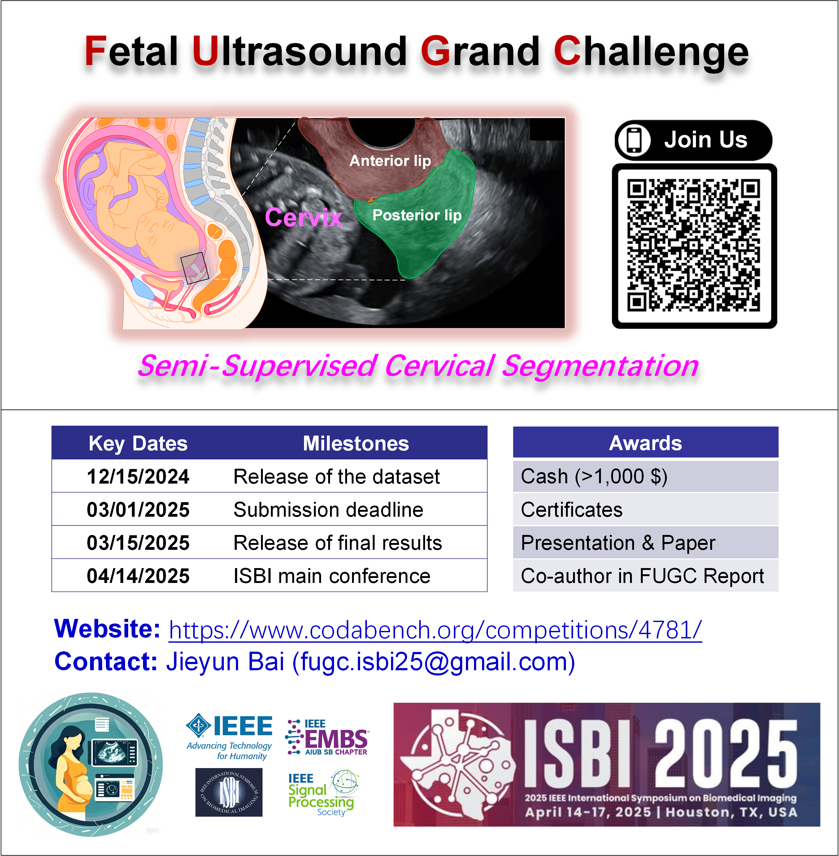
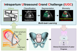
We extend the MICCAI PSFHS 2023 Challenge and the MICCAI IUGC 2024 Challenge from fully supervised settings to a semi-supervised setting that focuses on how to use unlabeled data.
I am an Associate Professor at Jinan University, a Joint Appointed Professor at the First Affiliated Hospital of Jinan University, and the Chief Scientist for Medical Artificial Intelligence at Guangzhou Lianyin Medical Technology Co., Ltd.
I was a postdoctoral researcher at the Auckland Bioengineering Institute, University of Auckland, working with Prof. Jichao Zhao from 2018 to 2019. I received my Ph.D. in Computer Science and Technology from Harbin Institute of Technology in 2017, under the supervision of Prof. Kuanquan Wang and Prof. Henggui Zhang, and I was also a visiting scholar at the Auckland Bioengineering Institute for one year.
My research focuses on AI-driven medical digital twins, ultrasound-based maternal–fetal monitoring, multimodal biomedical signal processing, and clinical decision support systems. I have published extensively on deep learning–based medical image and signal analysis in leading international conferences, including MICCAI, ISBI, EMBC, BIBM, and CinC, as well as in top-tier journals such as Medical Image Analysis (MIA), IEEE Transactions on Medical Imaging (TMI), IEEE Journal of Biomedical and Health Informatics (JBHI), and PLOS Computational Biology.
My team is dedicated to translational and data-driven medical AI research, leveraging large-scale clinical data collected from multi-center hospital systems. We actively develop open datasets, benchmark challenges, and open-source platforms to support reproducible and clinically relevant AI research.
I am a leading organizer in the international ultrasound AI community and currently serve as an Area Chair for MICCAI and ISBI Challenges. Through these initiatives, I have coordinated global collaborations among academia, hospitals, and industry, and have released large-scale, clinically curated datasets and standardized evaluation platforms to the community.
We are strongly committed to open science and have released more than 10 public datasets and code repositories on platforms such as Kaggle and Zenodo.
My group is currently recruiting postdoctoral fellows, research scientists, and graduate students in medical image analysis, medical digital twins, and AI-enabled maternal and cardiovascular healthcare.
Researchers and clinicians interested in medical AI and translational biomedical engineering are warmly welcome to contact me.
Website: https://jieyunbai.github.io/


Please sign in or register for FREE
If you are a registered user on Research Communities by Springer Nature, please sign in