From Space to Time: Live Tissue Profiling Using Nanoneedles
Published in Bioengineering & Biotechnology, Materials, and Protocols & Methods
Spatial omics is changing how we explore biology—giving us a map of where molecules are in tissues, and how they are organized. From measuring RNA in single cells1-4 to imaging proteins and metabolites across organs5, 6, these technologies have deepened our understanding of how tissues function in health and disease. But there’s a catch: almost all these tools work on fixed or frozen tissue slices. That means we are still looking at biology in a single moment in time—a snapshot, not a movie.
Scientists have long wanted to capture the dynamic nature of living systems, to watch molecular changes unfold in real time. A few platforms have tried to tackle this by sampling live cells at different time points, using fluidic force probes7 or advanced microscopy8. But these tools are often limited to cell cultures in dishes, and struggle when it comes to capturing the complexity of real tissues.
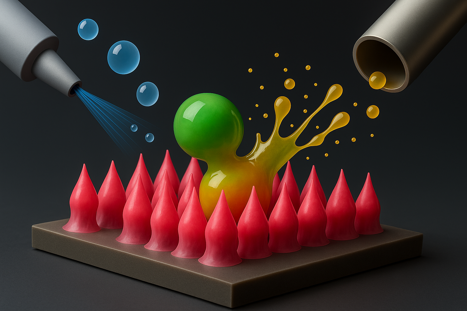
That’s where we thought of nanoneedles. These tiny, high-aspect-ratio structures can gently interact with cells and tissues, temporarily opening small pores in cell membranes to allow access without killing the cells (Figure 1). With the right chemistry on their surface, nanoneedles can extract a variety of molecules from inside living cells. And because they don’t cause lasting damage, they can return again and again to the same spot—like taking molecular snapshots across time9, 10.

Our group has spent years developing nanoneedles to monitor things like cellular energy (ATP)11, acidity (pH) 12, and enzyme activity in living cells13. And we are not alone in this effort. Across the field, scientists have been pushing the boundaries of what can be achieved with live-sample analysis. Porous silicon, for example, made it possible to gently extract molecules from live marine animals, helping to map their distribution14. Others have used diamond-based nanoneedles to capture genetic material directly from fresh tissues15. These advances all point toward a shared goal: to explore living systems without disrupting them. Yet, capturing how molecular landscapes evolve over time—what we might call "temporal omics"—is still a largely unexplored frontier. That’s the question we began asking: could we push further, into the dynamic world of living tissues, and bring both space and time together in one platform?
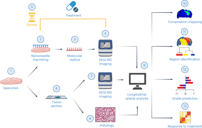
Our first breakthrough came when we successfully generated a nanoneedle imprint of a frozen mouse brain. The result was surely assisted by our department. Instead of sacrificing dedicated mice, we could coordinate with the other groups, to limit the animal culling and make use of animal tissue that would have been otherwise discarded. This collaboration allowed us to test extensively different strategies for the nanoneedle imprinting. Overall, the molecules onto the nanoneedles were the same observed in the original tissue, and we were able to obtain accurate maps of the frozen mouse brains. A crucial question was still open: is the spatial distribution of the molecules faithfully harvested onto the nanoneedles?
We chose DESI mass spectrometry to image the molecular maps. With DESI, a beam of charged droplets hits the nanoneedles, ionises the molecules harvested onto them, and delivers such molecules in secondary droplets towards a mass spectrometer. The choice of DESI was educated by studies suggesting synergy with the material forming the nanoneedles16, and by undergoing efforts in combining the technique in multimodal settings, for example by performing optical spectroscopy at the same time17. By scanning with DESI, we could confirm spatially that the nanoneedle imprinting generated an accurate molecular map.
Later, we learned how to generate an imprint from live brain slices. New questions quickly followed: would the cells survive the nanoneedles interfacing? Would they remain viable after ex-vivo culture? Weeks later, the data spoke for themselves: yes, they survived. That was our moment. If the tissue remained “happy” after one round of sampling, we knew we could do it again.
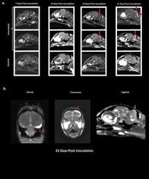
Glioma—a type of aggressive brain tumor—soon became our model system. These tumors drastically rewire how they use fats and lipids to survive and grow18. Their molecular makeup changes over time and space19, 20, making them an ideal target for our nanoneedle-based "spatiotemporal lipidomics". Our collaboration with Leor from Zaritsky’s lab at Ben-Gurion University brought a powerful layer of computational insight to the project. Together, we identified key lipid markers associated with glioma grade classification, transforming dense molecular maps into clinically meaningful patterns. In a way, this work reflects the art of bioengineering: combining intuition, creativity, and technical rigor to sculpt data into something more—something that tells a story about life in motion21.
Finally, like all good stories, further adventures await. Could we engineer nanoneedles for in-situ analysis? Could we combine lipid profiling with other molecular readouts22? And what other diseases might benefit from this approach?
We don’t have all the answers yet. But that’s what makes science so thrilling.
References:
- P. L. Ståhl et al., Science 353, 78-82 (2016).
- C. R. Merritt et al., Nature Biotechnology 38, 586-599 (2020).
- S. He et al., Nature Biotechnology 40, 1794-1806 (2022).
- K. H. Chen et al., Science 348, aaa6090-aaa6090 (2015).
- T. Guo et al., Nature 638, 901-911 (2025).
- N. M. Morato et al., Accounts of Chemical Research 56, 2526-2536 (2023).
- W. Chen et al., Nature 10.1038/s41586-022-05046-9 (2022).
- F. Marcuccio et al., Science Advances 10 (2024).
- C. Chiappini et al., Nature Protocols 16, 4539-4563 (2021).
- R. Elnathan et al., Nature Reviews Materials 7, 953-973 (2022).
- H. Kim et al., ACS Applied Materials & Interfaces 15, 49964-49973 (2023).
- C. Chiappini et al., ACS nano 9, 5500-5509 (2015).
- C. Chiappini et al., Advanced materials 27, 5147-5152 (2015).
- M. Ronci et al., Analytical Chemistry 84, 8996-9001 (2012).
- K. Xie et al., ACS Nano 15, 4881-4892 (2021).
- N. V. Schwab et al., Analytical Chemistry 86, 11722-11726 (2014).
- M. Jensen et al., Advanced Science 11 (2024).
- R. C. Gimple et al., Cancer Discovery 9, 1248-1267 (2019).
- M. S. Bergholt et al., ACS Central Science 4, 39-51 (2018).
- A. K. Jarmusch et al., Proceedings of the National Academy of Sciences 113, 1486-1491 (2016).
- N. H. Voelcker et al., Nature Reviews Bioengineering, (2025).
-
J. Bischof et al., npj Imaging 2, 5 (2024).
Follow the Topic
-
Nature Nanotechnology
An interdisciplinary journal that publishes papers of the highest quality and significance in all areas of nanoscience and nanotechnology.


Please sign in or register for FREE
If you are a registered user on Research Communities by Springer Nature, please sign in
Here are additional voices about the story from King’s College London.