Generating Synthetic Material Microstructures using Generative Adversarial Networks (GAN)
Published in Electrical & Electronic Engineering

To read our paper published in Scientific Reports, click here.
Material scientists conduct computer simulations to discover the linkage between microstructural morphology and material properties. In particular, for design and engineering of energetic materials (a term that includes propellants, explosives, and pyrotechnics), computational simulations performed on microscopic images of these materials are necessary to understand the mechano-chemical reactions which can flare up in a few nanoseconds. Energetic materials are typically mixtures of organic crystals, plasticizers, metals, and other inclusions, and these simulations illuminate how their complex micromorphology affects the sensitivity of such materials.
The problem, however, is that current manufacturing processes for energetic material offer little control over the microstructure morphology. This presents a considerable bottleneck to material scientists who desire to precisely control the response of energetic materials to external stimuli; the cut-and-try approach to materials design is extremely expensive and hazardous. Virtually prototyping of energetic materials with precise and predictable sensitivity requires conducting numerous simulations on a large collection of microstructures to accommodate the stochastic nature of the micromorphology. Unfortunately, imaged microstructure samples for a wide class of materials are not in abundant supply. The research community has found a get-around by developing techniques to create synthetic microstructures; traditionally this has been done by packing geometric primitives (circles, polygons, etc.) that are representative of the real morphology. However, their fidelity to the actual microstructures is questionable as they simplify the morphology; furthermore, packing objects at high densities can be challenging.
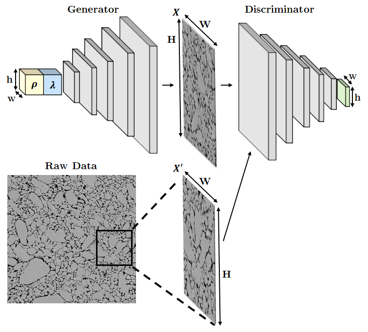
In our paper, newly published in Scientific Reports, we propose a novel method for generating synthetic microstructures using generative adversarial networks (GAN). Conceptually, GAN leverages a “competition” of two artificial neural networks, namely a Generator and a Discriminator—the generator generates a synthetic image while the discriminator attempts to distinguish synthetic images from real images. Interestingly, through such a competition, a generator learns how to generate synthetic images realistic enough to deceive the discriminator. Building upon this concept, we present a novel GAN architecture in our paper, which can generate synthetic microstructure images from parameterized inputs (Figure 1); there is no limit to the packing fraction for solids in our approach.

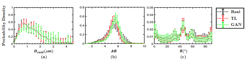
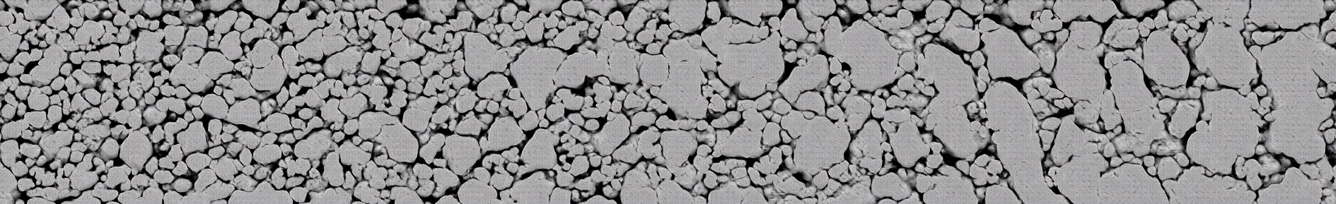
Two different types of parameters are used in our GAN model, global morphology parameters λ and local stochasticity parameters, ρ. Global morphology parameters define the overall “style” of the generated image. Local stochasticity parameters add natural stochastic variations to the morphology. Figure 2 shows microstructure images of cyclotetramethylene-tetranitramine (HMX) generated by the GAN model, whose difference from the real microstructure images is not discernable. Morphometry analysis in Figure 3 also suggests the realism of the GAN-generated microstructures. Moreover, this new GAN model can control and manipulate the microstructure morphology. For example, the animation in Figure 4 illustrates how micromorphology changes as the global morphology parameters of GAN are varied smoothly. This capability to smoothly vary the generated microstructures can be used, for example, to control the spatial layout of microscale morphology as seen in Figure 5.

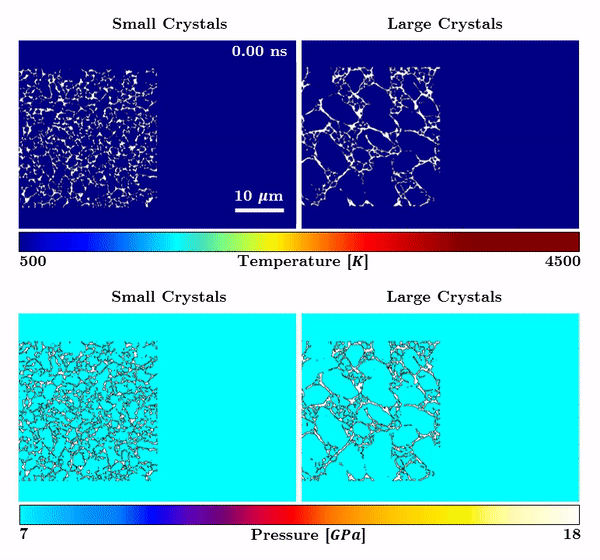
For a materials scientist, our work can be an exciting new tool to conduct morphology-controlled experiments to better understand structure-property linkages. For example, Figure 6 shows an animation of computer simulated shock-induced chemical reactions in two different GAN-controlled microstructures—one with smaller crystals/voids and another with larger crystals/voids. The sensitivity of the two cases are clearly different. For example, the temperature field for the sample with smaller crystals/voids (left column) shows a higher density of hot spots in the control volume when compared to the sample with larger crystals/voids (right column). Also, noticeable difference is seen in the pressure field between the two microstructures; similar to the hot spot temperature field, it is observed that the pqqressure field in the case of the small crystal size sample (left column) achieves higher overall magnitude in a larger part of the domain when compared to the pressure field for the sample with larger crystal. In future work, we will use these new capabilities to conduct controlled experiments and to quantify energetic material response as a function of morphology parameters. This will be significant step forward in energetic material research and will open new possibilities to “design” an energetic material with tailored property/performance characteristics.
This work was supported by the U.S. Air Force Office of Scientific Research (AFOSR) Multidisciplinary University Research Initiative (MURI) program (Grant No. FA9550-19-1-0318; PM: Dr. Martin Schmidt, Dynamic Materials program). We thank Drs. Chris Molek and Eric Welle from the Air Force Research Lab, Eglin AFB, for providing the images of pressed energetic materials; the images were obtained by Dr. Ryan Wixom at Sandia National Laboratories.
We are always looking for highly motivated and friendly PhD students and postdoctoral scholars eager to work on challenging scientific projects. Interested individuals are strongly encouraged to contact with Prof. Stephen Baek (stephen-baek@uiowa.edu). For more information, please visit our website.
Follow the Topic
-
Scientific Reports
An open access journal publishing original research from across all areas of the natural sciences, psychology, medicine and engineering.

Related Collections
With Collections, you can get published faster and increase your visibility.
Infectious disease diagnostics
Publishing Model: Open Access
Deadline: Sep 23, 2026
AI in Education
Publishing Model: Open Access
Deadline: Oct 09, 2026

Please sign in or register for FREE
If you are a registered user on Research Communities by Springer Nature, please sign in