How the brain corrects movements: inside our experiment that tested vision and proprioception during mid-reach perturbations
Published in Neuroscience
What we wanted to know
We have long known that both vision and proprioception (the body’s sense of limb position) guide reaching. But the PPC is a big square with different bars! Is it possible that some parts care more about vision while others care more about proprioception? That distinction matters: it tells us how flexible and optimized our brain can be when it has to switch strategies depending on what kind of sensory surprise happens. Understanding this could help design better neurorehabilitation protocols and prosthetic systems.
What did we do (in practice)
We asked volunteers to reach to touch a dot on a screen. In most trials nothing weird happened, just a normal reach to a target. But in rare cases (10% each) we either induce an unexpected correction of the movement by:
- Made the visual target suddenly jump.
- Pulled the participant’s wrist away from the target.

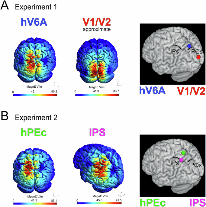
During the movements, we delivered transcranial magnetic stimulation to specific PPC sites to create a brief, reversible interference of neural processing. We targeted two medial PPC areas: hV6A (thought to be visually tuned) and hPEc (thought to be more somatosensory/proprioceptive), plus active controls and a ‘no-stimulation’ condition. If stimulating an area made corrections impaired, we can say that the area causally contributes to that correction.

Fig. 2 Localization of areas hV6A and hPEc on the brain.
How we measured “reaching corrections”
We tracked the trajectories of the index finger, the wrist, the forearm and the upper arm with a motion capture system and computed the 3-D Euclidean distance between perturbed and corresponding unperturbed trajectories over time. Smaller or delayed differences after stimulation compared with the ‘no-stimulation’ condition revealed an impairment in online correction.
So, what did we find?

Fig. 3 Simplified schematization of our scientific question. Modified image generated by ChatGPT.
hV6A is needed for rapid corrections when the target moves, while hPEc is needed for corrections when the arm is tugged. In other words: vision-driven fixes rely on hV6A; proprioception-driven fixes rely on hPEc. The PPC isn’t doing one generic “fix” job, because we found that its subregions specialize depending on the sensory source of the error.
The beauty of corollary findings
We didn’t just find the above-mentioned results, some other outcomes were delightfully interesting.
Stimulating hV6A reduced the mid-flight divergence (meaning that it delayed the visual correction), especially for the index, wrist and forearm, while the upper arm showed effects later in the movement, perhaps reflecting awkward postural compensation. Meanwhile, stimulating hPEc produced larger deviations during the proprioceptive perturbation, but the index finger was oddly resilient, likely because people used wrist/arm compensations to keep fingertip accuracy. We also saw that the stimulation of one of the control areas, namely the occipital cortex, impaired corrections to the proprioceptive pull, a curious hint that even early visual cortex can be engaged by somatosensory-related motor control!
Why it matters (in real life terms)
If different PPC subregions specialize for different sensory errors, therapies and brain-interface systems can be more targeted. For instance, for patients with proprioceptive loss (for example stroke recovery patients), stimulating or training circuits like hPEc may better restore limb stability than a one-size-fits-all approach. Neuroprosthetics and decoding algorithms could weight visual versus proprioceptive inputs differently depending on which cortical nodes are available or intact. More broadly, our study clarifies a neat principle: the brain routes optimize and pick the circuit best suited to the kind of information it senses.
Behind the scenes
Our participants sat through long sessions (about 3 hours) while we repeatedly disturbed their reaching movements with perturbations, and their arm was full of taped markers and electromyography patch electrodes. We want to deeply thank them for their resilience and the time they dedicated to science!
New Communications Biology paper: Functional Specialization of the Human Posterior Parietal Cortex in Visually and Proprioceptively Driven Reaching Corrections, by Brandolani R., Galletti C., Di Gloria D., Fattori P., Breveglieri R.
cognitive neuroscience, electrophysiology, transcranial magnetic stimulation
Follow the Topic
-
Communications Biology
An open access journal from Nature Portfolio publishing high-quality research, reviews and commentary in all areas of the biological sciences, representing significant advances and bringing new biological insight to a specialized area of research.

Related Collections
With Collections, you can get published faster and increase your visibility.
Artificial Intelligence Methodology in Structural Biology
Publishing Model: Hybrid
Deadline: Nov 30, 2026
Healthy Aging
Publishing Model: Open Access
Deadline: Dec 31, 2026


Please sign in or register for FREE
If you are a registered user on Research Communities by Springer Nature, please sign in