
Electron microscopy has reached atomic resolution, yet imaging hydrated samples in situ remains a major challenge. Water evaporates under high vacuum, and electrons induce radiolysis, leading to sample damage. Acquiring tilted image series to characterise the sample in 3D adds an extra level of complexity.
To bridge an existing gap in the field, we developed environmental liquid-phase electron tomography, capable of reconstructing hydrated samples in 3D at near-nanometer resolution while carefully controlling the electron dose to minimize beam-induced effects.
Overcoming Key Challenges
To achieve this, we had to overcome five simultaneous challenges:
- maintaining a stable hydrated state in environmental mode inside the electron microscope,
- ensuring sample alignment during tilt-series acquisition and optimizing the 3D reconstruction workflow,
- quantifying and controlling the deposited electron dose,
- achieving high spatial resolution appropriate to the sample,
- reaching sufficient temporal resolution so that the sample neither drifts nor degrades during acquisition.
Our initial focus was on environmental scanning electron microscopy (ESEM), with the longer-term goal of transferring the approach to environmental transmission electron microscopy (ETEM). ETEM offers complementary advantages, particularly in achievable spatial resolution.
Building the Tools
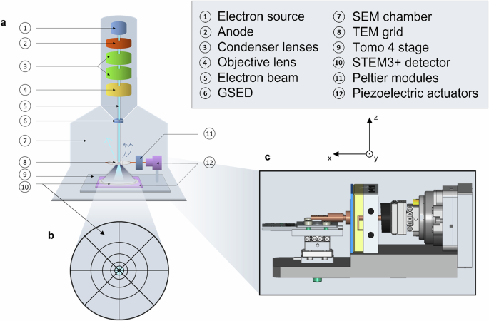
Since no commercial solution existed, we designed and built our own. On the hardware side, we developed a custom tomography stage with piezoelectric actuators and temperature control. On the software side, we created new algorithms to automatically align samples and predict drift before it occurred. These were integrated into a dedicated Python package, which allowed automated control of both the tomography stage and the microscopes for tilt-series acquisition.
With this integrated setup, we could collect dozens to hundreds of projections within minutes rather than hours—keeping acquisition times within the stability window of hydrated, beam-sensitive samples.
 column with the sample stage inside. b. Representation of the electron detector for versatile acquisition modes. c. Visualization of the home-made tomographic stage “Tomo 4”, comprising piezo-inertial elements and Peltier modules.")
Applications to Sensitive Materials
We first applied the method to aluminum hydroxide hydrogel, a medically relevant material. Using ESEM, we tracked hydration and dehydration cycles, while ETEM enabled us to quantify pore structures and nanoparticle distributions in 3D—without exceeding critical dose thresholds.
We also imaged unfixed, hydrated magnetotactic bacteria, which naturally produce intracellular iron oxide nanoparticles. This demonstrated the capability to study biological systems in near-native hydrated states, marking a step forward for both biology and nanoscience.
Closing Thoughts
By combining custom hardware, advanced algorithms, and optimized protocols, we demonstrate a practical route to 3D imaging of hydrated, beam-sensitive samples at near-nanometer resolution under controlled environmental conditions. With further improvements in hardware stability, this capability is expected to expand even more, opening new opportunities for nanoscale imaging of soft and biological matter.
Follow the Topic
-
Communications Engineering

A selective open access journal from Nature Portfolio publishing high-quality research, reviews and commentary in all areas of engineering.
Related Collections
With Collections, you can get published faster and increase your visibility.
Applications of magnetic particles in biomedical imaging, diagnostics and therapies
Publishing Model: Open Access
Deadline: May 31, 2026
Integrated Photonics for High-Speed Wireless Communication
Publishing Model: Open Access
Deadline: Mar 31, 2026


Please sign in or register for FREE
If you are a registered user on Research Communities by Springer Nature, please sign in