Intraoperative real-time blood monitoring
Published in Bioengineering & Biotechnology

Why do we need such a disruptive technology? Anticoagulant and thrombolytic therapy are widely used in the operating room, catheterization lab, and in the intensive care unit in order to reduce the risk of clotting, or to treat thrombosis events. However, whereas reducing the risk of thrombosis, anticoagulation therapy simultaneously increases the patient’s propensity to develop bleeding complications. Clinicians, therefore, must repeatedly adjust and maintain the dose of anticoagulant at the lowest possible level to prevent clot formation while simultaneously minimizing the risk of bleeding. To ensure that a surgical patient receives the correct dose of anticoagulant, clinicians must accurately assess the patient’s hemostatic status before, during, and after the surgical procedure. The need for strict monitoring also stems from the fact that patients often respond differently to identical anticoagulants. This is particularly true for heparin, which continues to be the most economical and widely used anticoagulant in the in-patient setting.
The current method of anticoagulation monitoring is labor intensive, expensive, and requires repeated withdrawal of blood from the circuit, thus exposing it to potential contamination. For these reasons, testing is performed only intermittently, resulting in 20–30 minute gaps in time when the precise status of anticoagulation is not known. Similarly, reversal of anticoagulation may be laborious and difficult to assess, as it relies on the same tests. What is needed are approaches that do not require specific sample collection and involved preparation, methods that are not based on end-point measurements, and techniques that can provide continuous real-time information. Our approach provides a viable solution.
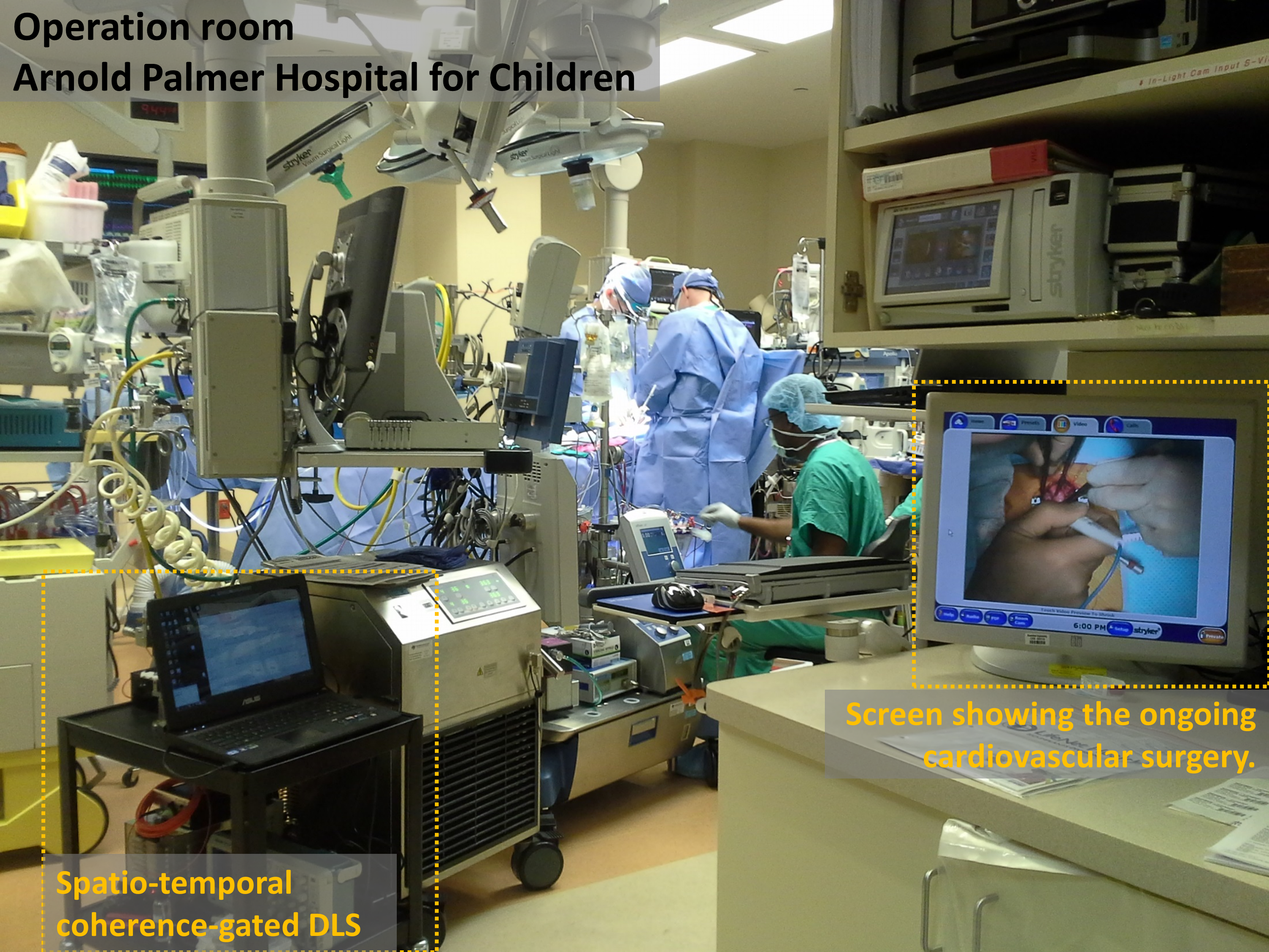
What are the specific technical challenges that we solved?Working with blood is complicated because of the high concentrations of scattering centers (red blood cells; RBCs), which may also be subject to additional advective flows and other variable environmental conditions that are specific to operation rooms. We solve this by using dynamic scattering of light, an approach that offers the possibility to have fast and nondestructive diagnostic tools. More specifically, we developed a robust implementation of a fiber-optic-based multimode common-path interferometer. The endoscopic nature of the measurement allows the sensor to be incorporated directly into standard vascular-access devices in medical equipment with minimal modifications and, therefore, to be safely operated in the operation room.

In addition to being robust, the sensing system must reliably provide information about blood dynamics over broad optical-scattering regimes without modifications to the hardware or the interpretation of the measurement. In the past this has been a notorious challenge. By three-dimensionally shaping the coherence properties of the illumination field, we have been able to assess blood coagulability independently of the scattering regime.
When I first came to Prof. Dogariu’s group I knew little about biomedical applications. I’ve been working in fiber-optic sensing but I was certainly not familiar with what it takes to bring fundamental physical concepts into actual medical applications. My first direct experience with translational research was really satisfying as I came to understand the value of thorough laboratory experiments and their role in making meaningful improvements to the measurement techniques. For instance, the steadiness of common-path interferometry, the optimal signal-to-noise balance afforded by the multimodal operation, and the effectiveness of statistical information retrieval are key features that allowed us to incorporate our optical sensor into the heart–lung machine in the operation room.
What was the evolution of this project? What is the relevance of this technology? The research in Prof. Dogariu’s group focuses on the interaction between light and complex media (http://random.creol.ucf.edu/). The lab was involved for a long time in developing optical-sensing techniques for characterizing various properties of complex fluids such as colloidal systems of relevance for pharmaceutical and microelectronics industries. The only fluid tissue in the body – blood – is also a complex fluid that can be thought as a highly-concentrated solution of non-spherical, soft particles. Hence, there was a natural transition to sensing the properties of biofluids. The work in Prof. Dogariu’s laboratory was initiated on the basis of cardiovascular programs funded by the Phillip Morris Research Foundation and the James and Esther King Biomedical Research Program.However, although our early laboratory and in vitro experiments showed great promise for this technology, we were not certain where to go next. The answer came when we met our co-investigator, William DeCampli, MD, Ph.D.He immediately recognized a particular potential application in his specialty, pediatric cardiac surgery.He acknowledged that the coagulation status of infants undergoing complex cardiac repairs can vary rapidly, putting infants (especially cyanotic infants) at risk for both thrombosis and bleeding during the conduct of the operation. Dr. DeCampli, who was an astrophysicist before becoming a cardiac surgeon, studied the physics and set up of the technique and realized that we could move directly to a clinical trial. We thought that if we could prove that our standard measures of clotting correlated closely with the continuously measured viscoelasticity index in a realistic setting, we would have a breakthrough in the management of these children, because we could detect changes early, in real time, and immediately see the effects of our interventions. Additionally, we would not need to draw blood repeatedly from the patient, as we do with the current standard methods of assessment.
Furthermore, because these types of optical measurements are fast and can be performed continuously, a number of different real-time monitoring applications could be enabled. For instance, such an optical instrument could also be used in a closed loop, for the automation of thrombolytic therapy delivery. In a ruggedized version, similar optical instrumentation could also be used in the field by first responders.
Our paper: Guzman-Sepulveda, J. R., Argueta-Morales, R., DeCampli, W. M. & Dogariu, A. Real-time intraoperative monitoring of blood coagulability via coherence-gated light scattering. Nat. Biomed. Eng. 1, 0028 (2017).
Images: Andrii Pshenychnyi.
![When PSMA-targeted therapy is not enough: high-risk localized prostate cancer after repeated [177Lu]Lu-PSMA radioligand therapy](/cdn-cgi/image/metadata=copyright,fit=scale-down,format=auto,quality=95,width=256,height=256/https://public-storage.zapnito.com/Ku6h7Yyp4Q0LXqRRMICCHR2v4LcOsmxMrmDPtOYuI1c)


Please sign in or register for FREE
If you are a registered user on Research Communities by Springer Nature, please sign in