Lighting the Path to Definitive Thyroid Cancer Detection
Published in Physics
The project started when Anna Yaroslavsky accidentally discovered that fluorescence polarization of methylene blue (MB Fpol) in cancerous skin is higher than in normal [1]. Later, Anna’s student, Rakesh Patel, measured higher MB Fpol in breast tumor cells from human clinical specimen [2]. To explain this finding, well-controlled cell culture studies with breast [3] and brain [4] cells, sponsored by the Massachusetts Technology Development Fund, were conducted by Anna and her student, Xin Feng. These studies revealed that MB Fpol is elevated in cancer due to greater uptake of positively charged MB in the negatively charged mitochondria of malignant cells. Other groups demonstrated that MB accumulation grows with increased mitochondrion membrane potential (MMP). Since increased MMP is a hallmark of many cancers, including brain, breast, thyroid, colon, renal, lung, and pancreatic cancers, MB Fpol may provide a quantitative method for diagnosing these cancers. Encouraged by cell culture research, together with clinical collaborators, Drs. Ashraf Khan, Lija Joseph, Andy Fischer, and Dina Kandil, Anna led two clinical trials with pathologically diverse cells acquired from cancer patients. These clinical investigations were conducted by Peter Jermain [5, 6]. While observing how Peter was adapting lengthy and sophisticated laboratory techniques for handling cell cultures to working with clinical specimens, Anna realized that clinical translation of MB Fpol technology hinged on developing a simple and rapid sample handling and data analysis techniques. That is when Anna met Prof. Thilo Stadelmann. Their complementary expertise led to a collaboration that yielded the results published here.
Fluorescence Polarization Imaging: A New Marker of Cancer
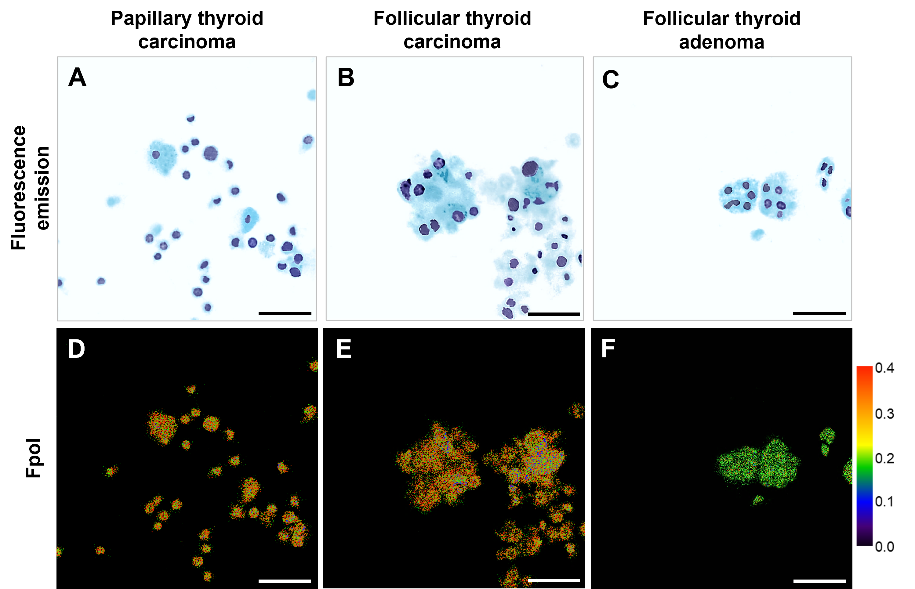
At the heart of the proposed approach lies the discovery that fluorescence polarization signal from MB is significantly higher in cancerous cells as compared to benign and normal cells. Fluorescence polarization is an optical technique that quantifies the degree of polarization of fluorescence light relative to the excitation light. Compared to other methods such as fluorescence emission and lifetime imaging, fluorescence polarization is less influenced by imaging protocols and more cost-efficient, respectively. In contrast to traditional methods that rely on visual assessments to identify abnormal morphological features of cancer cells MB Fpol provides a quantitative indicator of malignancy. Figure 1 illustrates the difference in Fpol for cancerous and benign cells.

Figure 1: MB-stained thyroid cells. The top row (A, B, C) shows fluorescence emission. The cells were pseudo-colored to mimic Papanicolaou stain. The bottom row (D, E, F) shows the same sample’s fluorescence polarization values. Panels D and F display malignant thyroid cells with higher Fpol values, whereas panel F shows benign thyroid cells with lower Fpol values. Adapted with permission from Reference 1/CC BY 4.0.
Automating AI-based Analysis for Speed and Accuracy
Manual analysis of Fpol images is labor-intensive and time-consuming. This is where Thilo’s students, Martin Oswald and Tenzin Langdun, stepped in. Through their award-winning bachelor thesis, they developed automated cell segmentation algorithm and obtained the first results showing significant time savings, as compared to manual processing. To prove that the automated approach reliably yielded the same results as manual, Ahmed Abdulkadir, spent countless hours with Peter, Santana, and Anna perfecting data processing approach and reanalyzing the original clinical data. Extensive validation showed that the diagnostic value obtained from the automated segmentation matched that of the manual segmentation (Figure 2).

Figure 2: (A) manually (MA) traced the cells; (B) automatically (AU) segmented cells. (C) Comparison between AU and MA methods showing difference in Fpol values versus difference in cell area. (D) Boxplot of AU and MA obtained Fpol.
The combination of Fpol imaging and AI is a potential game-changer to standard of care, offering several distinct advantages in thyroid cancer diagnostics:
- Objective Quantification: Fpol imaging provides a numerical value directly correlated with malignancy, eliminating subjective interpretation of cell morphology.
- Increased Efficiency: AI-powered automation streamlines analysis, enabling rapid and accurate diagnosis by decreasing the time it takes to evaluate a sample from hours to seconds.
- Enhanced Accessibility: Fpol method is more time- and cost-effective than competing technologies (e.g. multiplex molecular genetic testing).
A Glimpse into the Future
Our international interdisciplinary team is currently working on combining the newly established rapid cell processing protocol with automated image analysis to enable accurate triaging of thyroid cancerous cells within one hour or less. We hope that in the short-term this work will enable rapid and definitive diagnosis of thyroid malignancy. The long-term goal is to shift the paradigm of cancer diagnosis from subjective assessments based on visual inspection, to objective quantitative measurements that would enable accurate detection of malignant cells offering a new tool for early detection, diagnosis, and treatment monitoring.
References:
-
Yaroslavsky AN, Neel V, and Anderson RR, “Fluorescence polarization imaging for delineating nonmelanoma skin cancers,” Optics Letters. 29(14), 2010-2012 (2004).
-
Patel R, Khan A, Wirth D, Kamionek M, Kandil D, Quinlan R, and Yaroslavsky AN, “Multimodal optical imaging for detecting breast cancer,” J. Biomed. Opt. 17(6), (2012).
-
Yaroslavsky AN, Feng X, Muzikansky A, and Hamblin M, “Fluorescence polarization of methylene blue as a quantitative marker of breast cancer at the cellular level,” Sci. Rep. 9(1), (2019).
-
Feng X, Muzikansky A, Ross A H, Hamblin M, Jermain PR, and Yaroslavsky AN, “Multimodal quantitative imaging of brain cancer in cultured cells,” Biomed. Opt. Express. 10(8), 4237 (2019).
-
Jermain PR, Fischer AH, Joseph L, Muzikansky A, Yaroslavsky AN. “Fluorescence Polarization Imaging of Methylene Blue Facilitates Quantitative Detection of Thyroid Cancer in Single Cells,” Cancers 14(5), 1339 (2022).
-
Jermain PR, Kandil DH, Muzikansky A, Khan A, Yaroslavsky AN. “Translational Potential of Fluorescence Polarization for Breast Cancer Cytopathology,” Cancers 15, 1501 (2023).
Follow the Topic
-
Scientific Reports

An open access journal publishing original research from across all areas of the natural sciences, psychology, medicine and engineering.
Related Collections
With Collections, you can get published faster and increase your visibility.
Reproductive Health
Publishing Model: Hybrid
Deadline: Mar 30, 2026
Women’s Health
Publishing Model: Open Access
Deadline: Feb 28, 2026


Please sign in or register for FREE
If you are a registered user on Research Communities by Springer Nature, please sign in