Novel mechanism for pore formation!
Published in Biomedical Research
While therapeutic approaches for targeting the large toxins exist, there is nothing approved by the FDA to block CDT toxicity. Thus, an understanding of how CDT works is needed, so we can block the toxicity it delivers to host cells. As CDT toxicity is in essence irreversible once the CDTa toxin component enters the host cell, the most promising therapeutic strategies involve preventing CDTa entry into host cells via CDTb. To do this, our approach involves state-of-the-art structural biology techniques that can visualize structural and dynamic properties of both CDT components at atomic resolution. These data include how CDTb changes its conformation within different physiologically relevant spaces within the host before, during, and after CDTa toxin component is delivered into host cells, resulting in their rapid demise.
As CDT is in the binary toxin family, comparisons to other family members, including the anthrax toxin, were pursued to provide strategies to prevent CDTa cell entry. One such mechanism is to block CDTa from interacting with the cell-binding and toxin delivery component, CDTb. Such a strategy is ongoing in our laboratory and involves blocking sites on the toxin involved in CDT complex formation. This can be done using small molecules and/or via site-specific antibodies (i.e., biologics) directed at the CDTa-CDTb protein-protein interface (PPI). Another strategy is to directly stop CDT from interacting with host cell receptors, which involves targeting the receptor binding domains on CDTb. A third method is to clog the CDTb-dependent CDTa delivery system by strategically blocking its ability to form pores in the host cell membrane. Interestingly, we have discovered a novel mechanism of action used by CDTb to interact with the membrane of a cellular host and form pores in host cells that is unlike that ever before known previously for a binary toxin, including the anthrax toxin, which was reported by Abeyawardhane et al (2025) in Communications Biology (2).
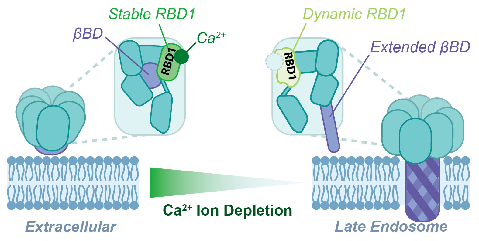
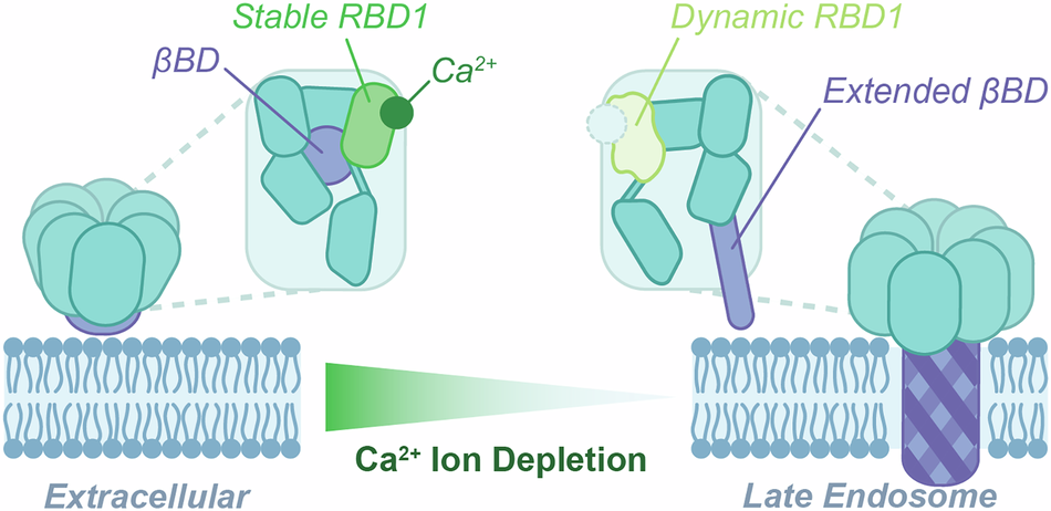
How was this discovery made and why did we have to wait so long to understand the new “trick” that c. difficile uses for entering and killing host cells? The bottom line up-front is that anthrax and CDT are both “activated” to change from a “protection” mode when they are outside the cell to an “attack” mode as they enter host cells via a normal cellular process termed endosomal delivery. In essence the toxins take advantage of normal changes in endosomal physiology that are important for the delivery of cargo used in normal cellular life. As it turns out, anthrax and several binary toxins takes advantage of a change in pH that lowers from neutral levels (pH = 7.4) to acidic levels (pH = 5.5). However, this was not the case for CDT, as lowering of pH did not enable the CDTb component of CDT to bind and form pores in lipid bilayers, as was found for anthrax. However, there is another large difference between conditions outside of a cell and within endosomes that were found to induce changes in CDTb conformation needed for it to move into its “host cell attack” mode. For CDT, it turned out to take advantage of a large decrease in Ca2+ concentration (>1000-fold change) upon entering endosomes rather than a pH change. Thus, when the Ca2+ levels were decreased to levels found in the endosome, the CDTb cell-binding and pore-forming component of CDT was found to “go into host attack mode”. Specifically, a newly discovered Ca2+-mediated trigger was discovered that involves dissociation of Ca2+ away from a domain in CDTb termed receptor binding domain 1 (RBD1). Upon Ca2+-release from RBD1, this domain becomes highly dynamic in nature, and it is these motions that instigate the forming of a “needle like” structure termed a “beta-barrel” that can bind and readily pierce lipid bilayers (Figure 1). This finding can now be incorporated into mechanistic studies involving how other physiologically important changes such as cellular pH change(s), host cell receptor binding, CDTa binding, and/or a combination of these events contribute to the structure and host cell toxicity from CDT. The results are also relevant to how CDTb alone can induce cellular inflammation, as was also reported recently for CDTb in the absence of CDTa (3).
- D. L. Abeyawardhane et al., The Importance of Therapeutically Targeting the Binary Toxin from Clostridioides difficile. Int J Mol Sci 22 (2021).
- D. L. Abeyawardhane et al., Pore formation by the CDTb component of the Clostridioides difficile binary toxin is Ca2+-dependent. Communications Biology 8, 901 (2025).
- K. Nabukhotna et al., Purified CDT toxins and a clean deletion within the CDT locus provide novel insights into the contribution of binary toxin in cellular inflammation and Clostridioides difficile infection. PLoS Pathog 20, e1012568 (2024).
Follow the Topic
-
Communications Biology
An open access journal from Nature Portfolio publishing high-quality research, reviews and commentary in all areas of the biological sciences, representing significant advances and bringing new biological insight to a specialized area of research.

Related Collections
With Collections, you can get published faster and increase your visibility.
Artificial Intelligence Methodology in Structural Biology
Publishing Model: Hybrid
Deadline: Nov 30, 2026
Healthy Aging
Publishing Model: Open Access
Deadline: Dec 31, 2026

Please sign in or register for FREE
If you are a registered user on Research Communities by Springer Nature, please sign in