The role of streptococcus lutetiensis in gastric cancer
Published in Cancer
Gastric cancer (GC) continues to be one of the most lethal cancers globally, and understanding the factors contributing to its development is crucial. Our recent studies have pointed to the microbiome, especially the presence of Streptococcus lutetiensis (S. lutetiensis), as a key player in gastric carcinogenesis. Here's what we know:
Key Findings
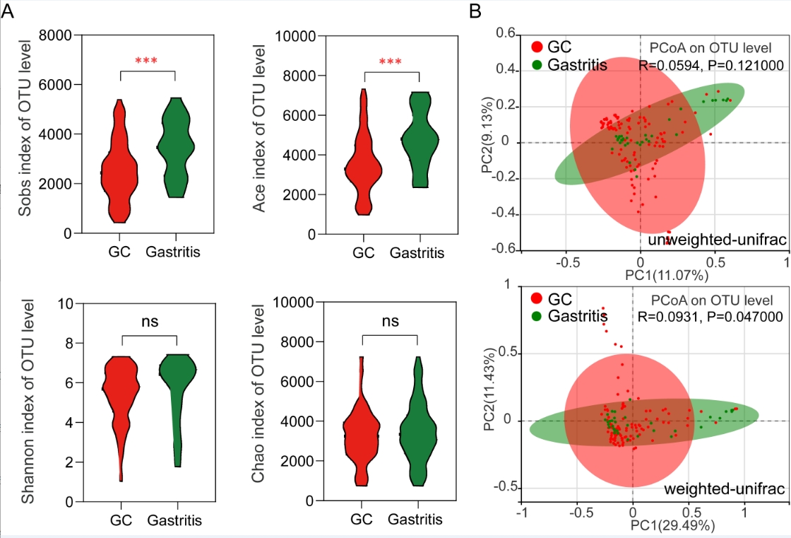
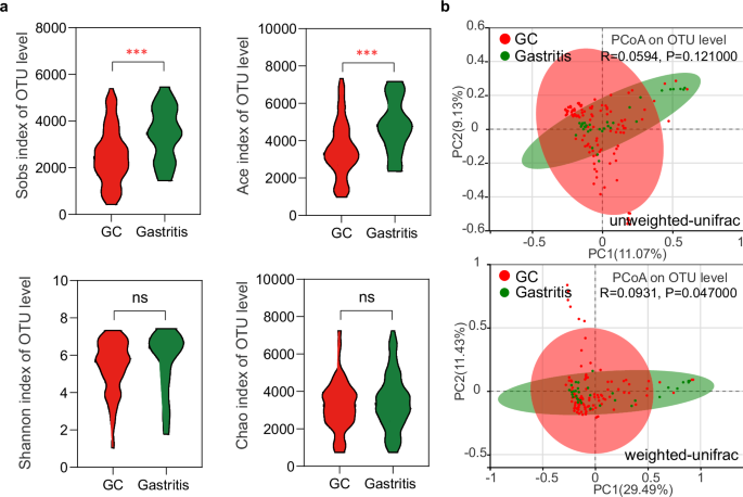
- Enrichment of lutetiensis in GC: Through advanced sequencing techniques, scientists have discovered that S. lutetiensis is significantly more abundant in the gastric tissue of cancer patients than in those with chronic gastritis. This bacterial species seems to contribute to the progression of gastric cancer.
- Impact on Immune Microenvironment: An intriguing finding from our study is the impact of lutetiensis on immune cells within the tumor microenvironment. Specifically, we focused on CD8+IL17A+ tissue-resident memory T (TRM) cells, which are key players against tumors. The presence of S. lutetiensis was found to significantly reduce the number of these immune cells, suggesting that this bacterium may hinder the immune system's ability to effectively combat cancer.
- Oxidative Stress and Immune Suppression: We discovered that S. lutetiensis activates oxidative stress responses in immune cells. This results in the impaired function of CD8+IL17A+ TRM cells, which produce key effector molecules like Granzyme A and IFN-γ to target and kill cancer cells. Essentially, S. lutetiensis is hijacking the immune response by activating stress pathways that diminish the effectiveness of these immune cells.
- Poor Prognosis Linked to High Levels of S. lutetiensis: High levels of S. lutetiensis in the gastric tumor environment correlate with more advanced stages of gastric cancer, indicating that this bacterium may serve as a marker for poor prognosis.
Why It Matters
This research highlights a significant, yet often overlooked, factor in cancer progression: the microbiome. S. lutetiensis doesn’t just hang around; it actively interacts with immune cells and can even manipulate the tumor microenvironment, making it more conducive to cancer growth. Understanding how S. lutetiensis operates within the immune system could open new doors for both diagnostic markers and potential therapies for gastric cancer.
Future Directions
The findings also prompt further exploration into how tumor bacteria influence cancer treatment. Could targeting S. lutetiensis help enhance immune responses in cancer therapies? Future research will likely investigate whether strategies that manipulate the microbiome could improve outcomes for patients with gastric cancer.
Follow the Topic
-
npj Precision Oncology
An international, peer-reviewed journal committed to publishing cutting-edge scientific research in all aspects of precision oncology from basic science to translational applications to clinical medicine.

Ask the Editor – Inflammation, Metastasis, Cancer Microenvironment and Tumour Immunology
Got a question for the editor about inflammation, metastasis, or tumour immunology? Ask it here!
Continue reading announcementRelated Collections
With Collections, you can get published faster and increase your visibility.
Minimal Residual Disease and Circulating Tumor DNA Dynamics in Personalized Cancer Treatment
Publishing Model: Open Access
Deadline: Mar 12, 2027
Demystifying Rare Breast Cancers
Publishing Model: Open Access
Deadline: Jul 16, 2026


Please sign in or register for FREE
If you are a registered user on Research Communities by Springer Nature, please sign in