The story behind the paper- An ALS assembly modulator signature in peripheral blood mononuclear cells: implications for ALS pathophysiology, therapeutics, and diagnostics.
Published in Chemistry and Biomedical Research

Background
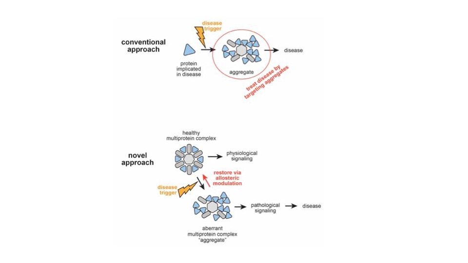
Homeostasis describes the healthy state. It is regulated by tightly controlled feedback loops which adjust in response to changes in the internal or external environment. While a lot has been learned about feedback and homeostasis over the last century, many feedback loops likely remain to be discovered – and we don’t know which are the most important ones. While a number of different triggering events can lead to disease, “sickness” can be fundamentally understood as departure from homeostasis. Likewise, manipulating feedback loops to restore homeostasis could treat currently incurable diseases such as ALS, a devastating neurodegenerative disease. Thus a fundamental goal of therapeutics ought to be restoration of homeostasis.
Unfortunately, it has not been known how to restore homeostasis. Therefore, most drugs today focus on targeting the cause of the disease – and hope that homeostasis will restore itself, once the disease is stopped. Usually it does, but sometimes, like the “colored wheel of doom” when your computer operating system is frozen, it does not – and it doesn’t work to try and undo the keystroke that caused your computer to freeze. As with your computer operating system, when that happens to homeostasis, you need a reboot. We hypothesized that ALS occurs when there is precisely such a failure to restore homeostasis after certain central nervous system injuries.
Enter viruses: they have been manipulating homeostasis by evolution through natural selection, most likely since the origin of life. They have figured out the best ways to disrupt homeostasis while silencing our defenses. If we could get them to tell us how they do that, and what those targets are, drugs with unprecedented therapeutic potential could be developed. That is what we believe we have done, and, courtesy of viruses, as described here, https://cassyni.com/events/88ZRW6kuvE9gZ8WXQPwJCm, our assembly modulator compound provides the necessary reboot of the operating system governing homeostasis, as it relates to ALS.
Our studies revealed first, that catalyzed protein assembly is a crucial weak link in biology that viruses exploit. In effect, we used viruses as “trufflehounds” to reveal those high value targets that are inaccessible to conventional drug discovery tools. For example, powerful tools such as CRISPR are unable to parse small subsets of a gene product that do different functions based on differences in protein-protein interactions, e.g. due to covalent post-translational modification, intrinsically unfolded domains or different pathways of biogenesis and assembly, all of which have been demonstrated to occur.
While advancing these drugs against viruses, we discovered that some of them also work on particular non-viral diseases. Thus, when we learned of work by others revealing a relationship between ALS and retroviruses (for example, HIV), we assessed our anti-retroviral assembly modulator compounds -- and found several of them active on cellular models of ALS. Advanced analogs not only became safer and more potent against ALS, but some actually lost their anti-viral activity, becoming specific for ALS.
In this paper, we sought to further explore the relationship between ALS-selective assembly modulator compounds and homeostasis using blood samples from ALS patients versus healthy individuals. The results are important not only for what they reveal about previously unappreciated feedback loops relevant to ALS, but also because they provide an extremely early signature of ALS by which it can be diagnosed prior to significant disability – using a drug that is strikingly therapeutic for ALS.
Key Findings- Identification of a protein signature in ALS patient PBMCs and a related feedback response upon treatment of ALS patient blood samples
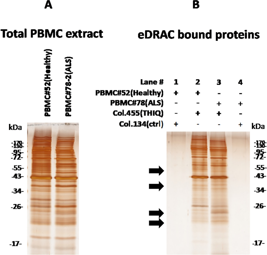
When ALS assembly modulator compounds are used as “fish hooks” to detect their targets, they bind to distinctly different protein complexes in PBMCs (white blood cells) from healthy vs ALS patients. The proteins RanGTPase and p62/SQSTM1 were found to have an inverse relationship in the small amounts found in the ALS drug target. P62 is a crucial regulator of autophagy (a fundamental process of host defense), and is lost from the drug target with disease progression. Ran GTPase regulates protein transport in and out of the nucleus, long suspected defective in ALS, specifically with regards to the protein TDP-43. A distinctive fragment of RanGTPase increases with ALS progression. Furthermore, our data reveals new feedback loops implicated in ALS upon which these compounds act.
Next Steps
This work provides foundation for important next steps, including development of an ALS therapeutic, an ALS biomarker, and a way to understand ALS as a disease of homeostasis.
ALS Therapeutic
The ex vivo response we observed is that when ALS patient blood samples are treated with the same compound that is efficacious in animal models, degradation of RanGTPase– likely through feedback– is reduced. Efficacy on a novel target in ALS patient blood, distinctive to ALS, and related to the target seen in ALS mouse brain where the compound is proven efficacious, is encouraging and mandates further exploration.
ALS Biomarker
This signature was present and detectable early in the course of ALS, including even before the patient was disabled. The observed ALS signature in blood could both enable a diagnostic test to detect ALS early, prior to disability, and provide a biomarker for ALS clinical trial design, monitoring and objective assessment. Any treatment for ALS will be more effective if it can be administered to rescue motor neurons before they die. This creates the possibility of a future where ALS patients can be treated early in the disease to prevent disability and achieve a cure.
Tools for Understanding ALS as a disease of homeostasis
ALS is viewed as a disease of motor neurons. However, our findings, derived from ALS patient blood, suggest ALS may actually be a systemic disease, manifest most severely in motor neurons. Further studies using the compounds as molecular probes could provide insight into ALS pathogenesis.
Conclusion
Drugs of the future must do more than just stop primary diseases. They need to restore homeostasis. Evidence, including some presented in this work, suggest assembly modulators do just that for ALS. We look forward to following up these studies, developing assembly modulators as diagnostics and therapeutics for ALS, and using them as tools to better understand the molecular basis for homeostasis, the best hope for superior ALS drugs in the future.
Follow the Topic
-
Clinical Proteomics
This journal encompasses all aspects of translational proteomics. This includes quantitative and qualitative profiling of proteins and peptides that are present in clinical specimens like human tissues and body fluids.

Related Collections
With Collections, you can get published faster and increase your visibility.
Biofluid Proteomics: Unlocking Molecular Insights from Body Fluids
Biofluid proteomics is an emerging and dynamic field within clinical proteomics that focuses on the comprehensive analysis of proteins present in biological fluids such as blood, urine, cerebrospinal fluid, saliva, and others. These fluids serve as accessible windows into physiological and pathological states, offering immense potential for biomarker discovery, disease monitoring, and personalized medicine. Advances in mass spectrometry, sample preparation, and computational tools have enabled high-resolution profiling of complex proteomes, driving translational research from bench to bedside.
This Collection aims to showcase cutting-edge studies that leverage biofluid proteomics to deepen our understanding of disease mechanisms, improve diagnostic accuracy, and identify therapeutic targets. We welcome contributions that span methodological innovations, clinical applications, and integrative multi-omics approaches. By gathering diverse perspectives, this collection seeks to accelerate the development of robust, reproducible, and clinically relevant proteomic strategies.
Topics of interest include, but are not limited to:
• Novel sample preparation and enrichment strategies for low-abundance proteins in biofluids.
• Mass spectrometry-based workflows for high-throughput and quantitative biofluid proteomic analysis.
• Biofluid-based biomarker discovery and validation for early disease detection and prognosis.
• Integration of proteomics with other omics (genomics, metabolomics) for biofluid-based systems-level insights.
• Clinical applications of biofluid proteomics in oncology, neurology, cardiovascular, and infectious diseases.
All submissions in this collection undergo the journal’s standard peer review process. Similarly, all manuscripts authored by a Guest Editor(s) will be handled by the Editor-in-Chief. As an open access publication, this journal levies an article processing fee (details here). We recognize that many key stakeholders may not have access to such resources and are committed to supporting participation in this issue wherever resources are a barrier. For more information about what support may be available, please visit OA funding and support, or email OAfundingpolicy@springernature.com or the Editor-in-Chief.
Publishing Model: Open Access
Deadline: Nov 16, 2026
AI Approaches in Clinical Proteomics: From Data to Discovery
Artificial intelligence (AI) is transforming clinical proteomics by introducing advanced computational capabilities that enable data analysis, pattern recognition, and predictive modeling for biomarker discovery and precision medicine. While AI encompasses a broad conceptual framework, this Collection emphasizes the practical application of AI capabilities within clinical proteomics workflows, such as machine learning, deep learning, and integrative computational tools.
We aim to showcase protocols, algorithms, and frameworks that leverage these capabilities to improve data interpretation, enhance reproducibility, and accelerate translational research. Contributions may include novel AI-driven models for protein expression profiling, multi-omics integration, disease classification, and therapeutic target identification. By bringing together experts in computational biology and clinical proteomics, this Collection seeks to foster innovation at the intersection of data science and molecular medicine.
Topics of interest (but not limited to):
• Machine learning and deep learning models for clinical proteomics data
• AI-driven biomarker discovery and disease prediction
• Integrative approaches combining proteomics with other omics (genomics, metabolomics)
• Data preprocessing, normalization, and feature selection for AI workflows
• Explainable AI and model validation in clinical research
All submissions in this collection undergo the journal’s standard peer review process. Similarly, all manuscripts authored by a Guest Editor(s) will be handled by the Editor-in-Chief. As an open access publication, this journal levies an article processing fee (details here). We recognize that many key stakeholders may not have access to such resources and are committed to supporting participation in this issue wherever resources are a barrier. For more information about what support may be available, please visit OA funding and support, or email OAfundingpolicy@springernature.com or the Editor-in-Chief.
Publishing Model: Open Access
Deadline: Oct 15, 2026

Please sign in or register for FREE
If you are a registered user on Research Communities by Springer Nature, please sign in