Unique digital dog and cat skull database
Published in Ecology & Evolution, Agricultural & Food Science, and Zoology & Veterinary Science

Tibor Csörgő, a researcher at ELTE, has been collecting animal skulls for decades to teach anatomy to biologists. The shape of the skull varies considerably between species and breeds, especially in dogs, where, for example, greyhounds have long skulls and the now popular French bulldogs have rounded skulls. A skull biobank could be a valuable resource for education, medicine and evolutionary research. For example, Zsolt László Garamszegi, Director of the HUN-REN Institute of Ecological Research, together with ethologists from ELTE and Niclas Kolm from Stockholm University, have based their findings in part on this collection, which shows that modern dog breeds bred in the last 200 years have larger brains than those with ancient origin, due to altered selection effects. The researchers wanted to make this unique collection of skulls available to all.
Similar research previously required researchers to visit collections in person. Today, however, it is possible to digitise skulls so that anyone can conduct studies at their desk, even on another continent. The digitisation was carried out by Kálmán Czeibert, a veterinary neuroanatomist in collaboration with Ádám Csóka, Tamás Donkó and Örs Petneházy, imaging specialists from the Medicopus Nonprofit Ltd. research unit, using a medical high-resolution computed tomography (CT) scanner. In total, 431 skulls were digitised, representing 152 dog breeds, 9 cat breeds and 12 of their wild relatives, including wolves, jackals, coyotes, a leopard and a serval.
The database was published in the journal Scientific Data. According to the study's corresponding author, Enikő Kubinyi, head of the MTA-ELTE Lendület Companion Animal and ELTE NAP Canine Brain research groups, "the digital skull database can be used for comparative anatomical and evolutionary studies, in the education of veterinarians and biologists, and even for the development of machine learning algorithms for automated species identification and veterinary diagnostics". The researchers have also produced a video to illustrate the database, which can be viewed here.
Original article: Czeibert, K., Nagy, G., Csörgő, T., Donkó, T., Petneházy, Ö., Csóka, Á., Garamszegi, L. Z., Kolm, N., Kubinyi, E. (2024) High-resolution computed tomographic (HRCT) image series from 413 canid and 18 felid skulls. Scientific Data, https://doi.org/10.1038/s41597-024-03572-x
Image: Skull of a Saint Bernard dog from different views. First row: right, left, and front views. Second row: top, bottom, and back views. Credit: Kálmán Czeibert
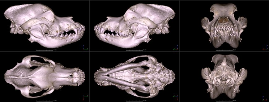
Image: Skull of a Boston terrier on a CT image series. First row: transverse and sagittal views. Second row: dorsal view. Credit: Kálmán Czeibert
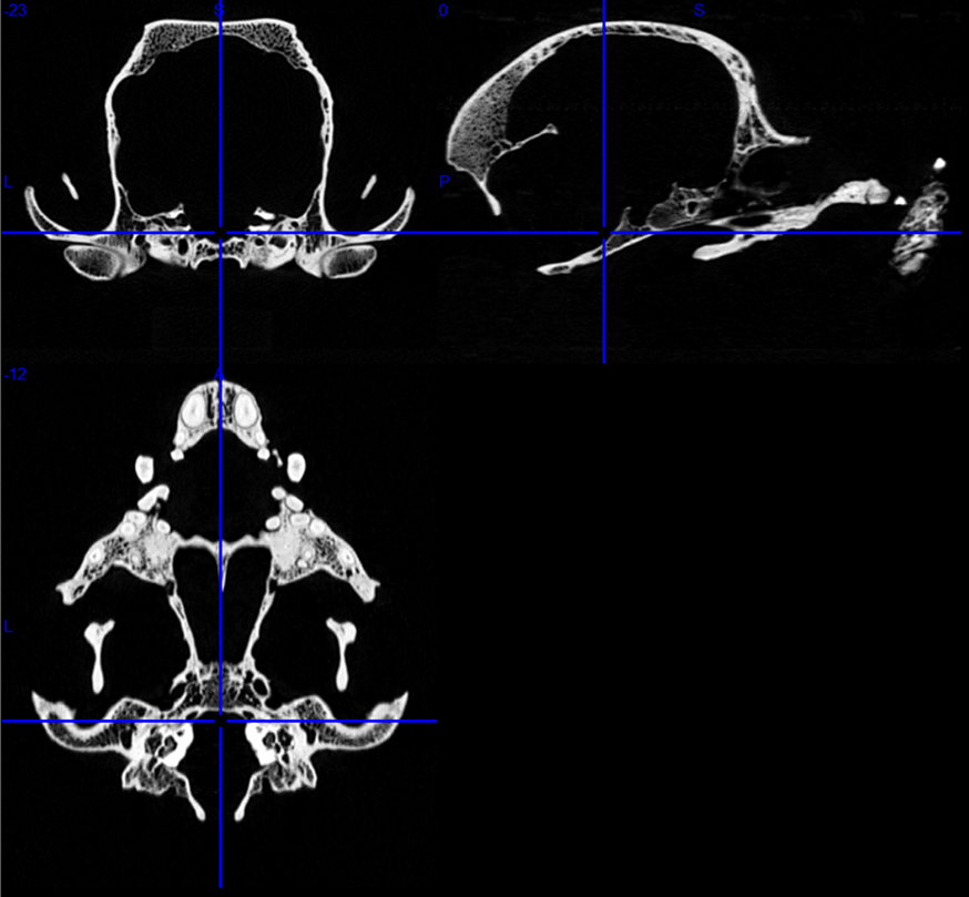
Image: Dolichocephalic (A), mesocephalic (E), and brachycephalic (I) dog skulls and their corresponding endocranial casts (brain models, B, F, J, respectively). The endocranial casts have been developed based on the digitised skulls. The middle ectosylvian auditory area is highlighted in red. Credit: Kálmán Czeibert
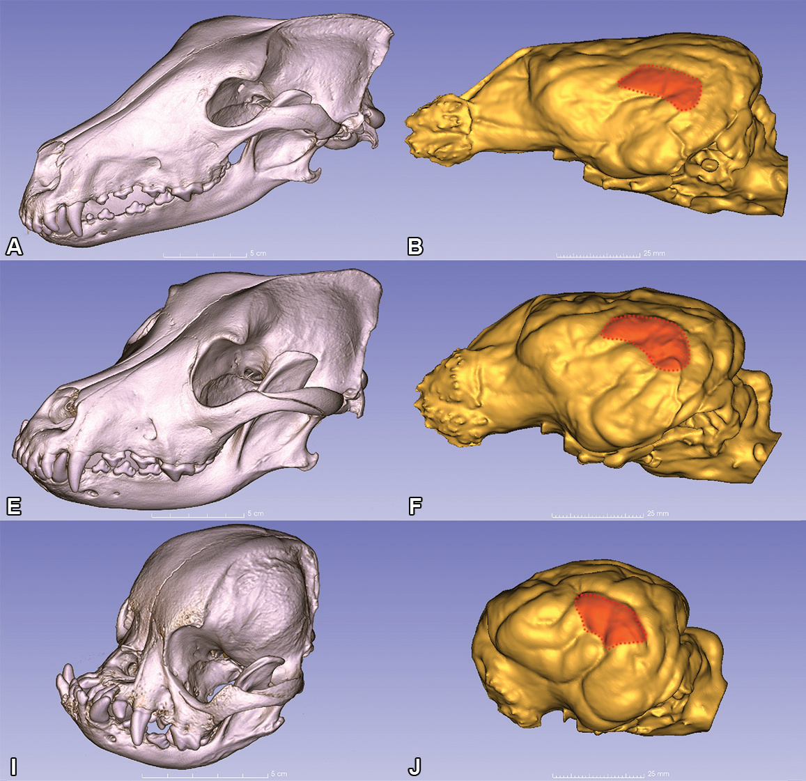
Image: Skulls at ELTE, Institute of Biology, Budapest (Credit: Eniko Kubinyi)
Funding: The study was supported by the Hungarian Academy of Sciences via a grant to the MTA-ELTE ‘Lendület/Momentum’ Companion Animal Research Group (grant no. PH1404/21), the National Brain Programme 3.0 (NAP2022-I-3/2022), the Hungarian Ethology Society, National Research, Development and Innovation Office (grant no. 2019-2.1.11-TÉT-2020-00109), the Ministry of Economy and Competitiveness in Spain (CGL2015-70639-P), and the Swedish Research Council (grant no. 2021-04476 and 2016-03435). Open access funding was provided by ELTE Eötvös Loránd University.
Follow the Topic
-
Scientific Data

A peer-reviewed, open-access journal for descriptions of datasets, and research that advances the sharing and reuse of scientific data.
Related Collections
With Collections, you can get published faster and increase your visibility.
Data for crop management
Publishing Model: Open Access
Deadline: Apr 17, 2026
Genomics in freshwater and marine science
Publishing Model: Open Access
Deadline: Jul 23, 2026

Please sign in or register for FREE
If you are a registered user on Research Communities by Springer Nature, please sign in