Video gaming facilitates adaptation to surgical exoscopes
Published in Surgery
Exoscopes in Neurosurgery
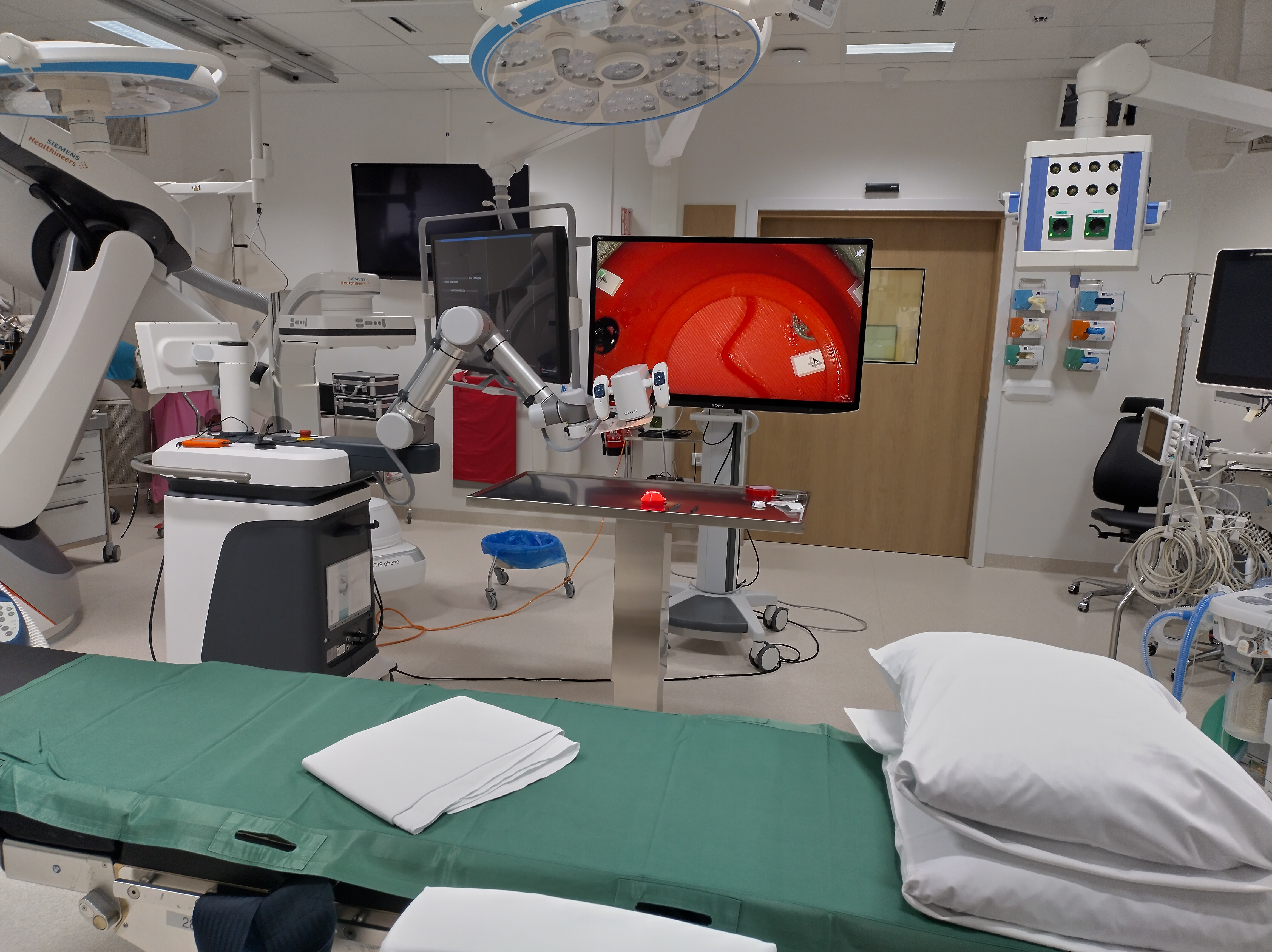
Three-dimensional (3D) digital exoscopes are novel tools for the magnification and visualization of structures in neurosurgery [1,3]. A robotic arm, equipped with a 3D digital camera, is controlled by a surgeon e.g., with a footpedal. The camera’s view of the surgical field is projected on a 3D monitor. Exoscope use in complex neurosurgery has led to outcomes comparable to the ones attained with a microscope [6,8]. Exoscopes allow surgeons to maintain an ergonomic posture during surgery, while enabling accurate target visualization and a wide range of motion [2,4,9].
Building a Microsurgical Training Model
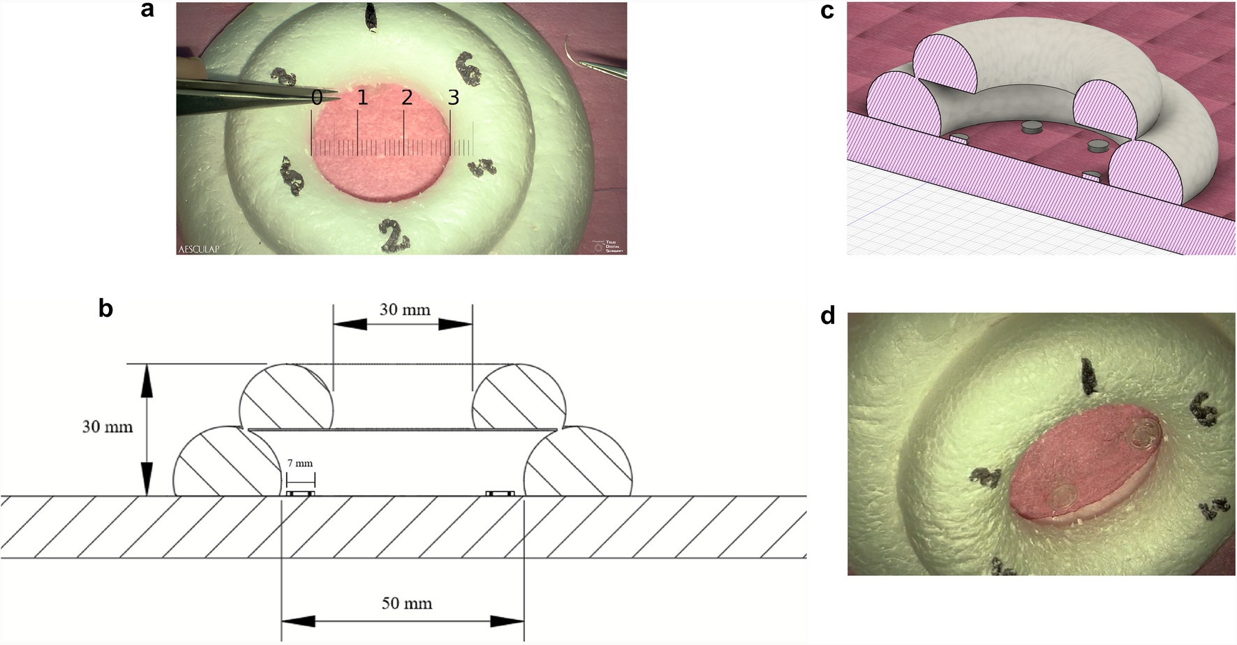
We developed a microsurgical training model for evaluating exoscope use. The model is represented in Figure 1.

Figure 1. a) The model from above [cm]. This image has been captured using the exoscope. b) A 2D cross section of the model. Created using Autodesk Fusion. c) A 3D cross section of the model. Created using Autodesk Fusion. d) An oblique view of the model, showcasing the normally concealed target discs. This image has been captured using the exoscope
The model consists of two Styrofoam rings forming an inverted funnel with the orifice smaller than the base. Inside, there are six symmetrically positioned plastic discs, 0.7 cm in diameter, attached to the base of the model. Each disc contains four symmetrically positioned slits, through which a microneedle has to be passed as shown in Video 1.
Recruiting Participants
We examined the impact of video gaming experience on performance, by recruiting 20 medical students. None had prior experience with exoscopes, microscopes, or any form of micro- or macrosurgery. We divided them into two groups:
- Gamers (n=11), who had more than 1,000 hours of video gaming.
- Controls (n=9), with less than 500 hours of gaming experience.
What We Found
Overall, students with gaming experience adapted to the exoscope faster. Gamers moved and tilted the exoscope camera less frequently, suggesting they needed fewer adjustments to get a clear view. Yet, despite fewer camera movements, they maintained similar zoom levels and target visibility as the control group.
When it came to speed, gamers completed all tasks faster—taking a median of 10 minutes 14 seconds compared to 13 minutes for the controls. This was largely due to the gamers using the exoscope more efficiently, since both groups took similar time to pass the needle through the target discs. However, both groups improved over time, underscoring the benefits of even short-term training.
Fine motor skills seemed similar. Both groups took about the same time to pass the needle through the slits and dropped the needle equally often. This suggests that gaming did not improve manual dexterity directly. A previous study also concluded that there was no correlation between video gaming or instrument playing and improved microsurgical performance in a group of 46 students [5]. Nevertheless, in our study, gamers drifted out of the surgical field with their forceps significantly less often than the control group, which may reflect superior 3D perception and spatial awareness.
It is possible that gamers simply adapt faster to the exoscope foot pedal, however, the results are probably best explained by superior 3D perception among the gamers, which results in better exoscope utilization. This is supported by the fact that while both groups employed similar zoom, the gamers exited the surgical field with their forceps less often than the controls.
A Note of Caution
We don’t believe video gaming alone makes a good surgeon. Microsurgery requires far more than fast adaptation to moving an exoscope camera. But our findings suggest that frequent exposure to immersive 3D environments—like video games—might ease the learning curve when adopting the exoscope and perhaps other new surgical technologies as well.
Looking Ahead
These results highlight an opportunity: virtual simulations could become powerful tools for surgical training in exoscopic neurosurgery. An accessible exoscope simulator with realistic foot pedal controls and 3D surgical models could help novices build confidence before stepping into the operating theatre.
Our team has begun developing such a simulator. We also believe that even brief, structured training—perhaps just six hours—can significantly improve performance and comfort with exoscopes [7].
As digital tools become integral to surgery, finding new ways to prepare and support learners will be essential. And the next generation of surgeons may already be honing some of their skills—without even realizing it—while playing video games at home.
References
- Langer DJ, White TG, Schulder M, Boockvar JA, Labib M, Lawton MT (2020) Advances in Intraoperative Optics: A Brief Review of Current Exoscope Platforms. Oper Neurosurg (Hagerstown) 19:84-93. doi:10.1093/ons/opz276
- Moisi MD, Hoang K, Tubbs RS, Page J, Fisahn C, Paulson D, Jeyamohan S, Delashaw J, Hanscom D, Oskouian RJ, Chapman J (2017) Advancement of Surgical Visualization Methods: Comparison Study Between Traditional Microscopic Surgery and a Novel Robotic Optoelectronic Visualization Tool for Spinal Surgery. World Neurosurg 98:273-277. doi:10.1016/j.wneu.2016.11.003
- Montemurro N, Scerrati A, Ricciardi L, Trevisi G (2022) The Exoscope in Neurosurgery: An Overview of the Current Literature of Intraoperative Use in Brain and Spine Surgery. Journal of Clinical Medicine 11:223
- Muhammad S, Lehecka M, Niemelä M (2019) Preliminary experience with a digital robotic exoscope in cranial and spinal surgery: a review of the Synaptive Modus V system. Acta Neurochirurgica 161:2175-2180. doi:10.1007/s00701-019-03953-x
- Osborn HA, Kuthubutheen J, Yao C, Chen JM, Lin VY (2015) Predicting Microsurgical Aptitude. Otol Neurotol 36:1203-1208. doi:10.1097/mao.0000000000000798
- Rossmann T, Veldeman M, Nurminen V, Huhtakangas J, Niemelä M, Lehecka M (2023) 3D Exoscopes are Noninferior to Operating Microscopes in Aneurysm Surgery: Comparative Single-Surgeon Series of 52 Consecutive Cases. World Neurosurg 170:e200-e213. doi:10.1016/j.wneu.2022.10.106
- Vasankari V, Hafez A, Pohjola A, Auricchio AM, Calvanese F, Rossmann T, Veldeman M, Badic I, Netti E, Rautalin I, Nurminen V, Raj R, Niemelä M, Lehecka M (2024) Even short-term training improves the skills of novice exoscope users: a prospective laboratory experiment. Acta Neurochirurgica 166:118. doi:10.1007/s00701-024-05975-6
- Veldeman M, Rossmann T, Huhtakangas J, Nurminen V, Eisenring C, Sinkkonen ST, Niemela M, Lehecka M (2023) Three-Dimensional Exoscopic Versus Microscopic Resection of Vestibular Schwannomas: A Comparative Series. Oper Neurosurg (Hagerstown) 24:507-513. doi:10.1227/ons.0000000000000602
- Zhang Z, Feng Y, Lu X, Yang B, Zhang H, Ma Y (2022) Microvascular anastomosis in a challenging setting using a 4 K three-dimensional exoscope compared with a conventional microscope: An in vivo animal study. Front Surg 9:1021098. doi:10.3389/fsurg.2022.1021098
Follow the Topic
-
Acta Neurochirurgica
This is a global journal focusing on clinical neurosurgery and relevant neuroscience research.

Related Collections
With Collections, you can get published faster and increase your visibility.
Contemporary Treatment of Skull Base Lesions
After the “glory days” of the 1990s—marked by progressively more extended approaches and the pursuit of ever more radical resections of skull base lesions, often at the expense of neurological function—the pendulum swung. Skull base surgeons, radiotherapists, neuro-otologists, and other ENT surgeons adopted more conservative strategies, with partial resections followed by adjuvant treatment becoming the norm. Gradually, the pendulum shifted again in an effort to balance progression-free survival with neurological preservation, leading to the contemporary emphasis on “maximal safe resection.”
Given the rapid evolution of the field, this topical collection brings together experts from around the world to discuss the current standards of care for a variety of skull base lesions. Contributions include state-of-the-art systematic reviews, clinical research studies, and innovative meta-analyses. “How I do it” submissions will also highlight new technical advances in skull base surgery.
This collection brings together international experts to review modern strategies for managing skull base lesions, emphasizing the balance between oncological control and neurological preservation. It showcases up-to-date evidence, evolving surgical techniques, and practical insights from leading skull base surgeons.
Publishing Model: Open Access
Deadline: Nov 01, 2026


Please sign in or register for FREE
If you are a registered user on Research Communities by Springer Nature, please sign in