Water-based non-toxic MRI contrast agents
Published in Chemistry
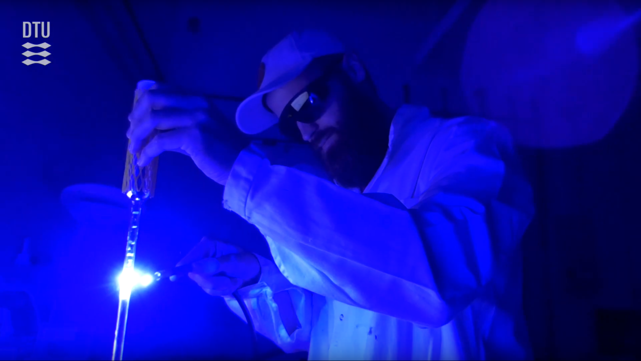
Despite all efforts and progress medicine and technology have made during the last decade, early and safe detection of a disease remains a powerful way to save lives. For instance, diagnosing a tumor before it progresses opens up for an increased number of available treatment options, increased survival rate and improved quality of life.
To this regard, Magnetic Resonance Imaging (MRI) techniques allow non-invasive spatial differentiation of flowing and static matter by using nonionizing radiofrequency radiation.1,2 Based on the physical principles of Nuclear Magnetic Resonance (NMR), the technique exploits the signal coming from the nuclear magnetism of water protons (1H nuclei) contained in the body’s soft tissues.
Although these techniques are currently used in clinical practice, there is still a need for higher reader sensitivity and enhanced contrast on images.3,4 Often, this is achieved by injecting gadolinium-based contrast agents that modify the magnetic properties of the water in specific areas of the body. Unfortunately, there are serious concerns about the safety of gadolinium-based contrast agents for certain groups of patients.5,6
A work around could be to generate ex-situ water gifted with high contrast and inject it into the body prior to examination.
Our group in Denmark is specialized in dissolution Dynamic Nuclear Polarization (dDNP),7 a technique invented in 2003 and able to increase NMR sensitivity in the liquid state by generating injectable solutions that are “hyperpolarized” (i.e. NMR signal 10000-fold higher than normal).
Among other topics, we have been focusing on hyperpolarizing water for MRI applications, targeting high-contrast perfusion (Figure 1) and angiographic imaging.
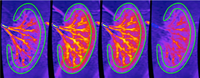
Figure 1: Renal angiography in a pig injected with water-based contrast agents at different time steps.8
However, hyperpolarizing water is far from trivial. This is mostly because of the paramagnetism of stable radicals used in the dDNP process to create the hyperpolarization: at the moment of injection they are the main source of signal, thus MR contrast, loss. Images in Figure 1 were recorded by perfusing a pig kidney with water polarized to 5% only. How beautiful would it be to record images using water hyperpolarized to its 100 % theoretical limit?
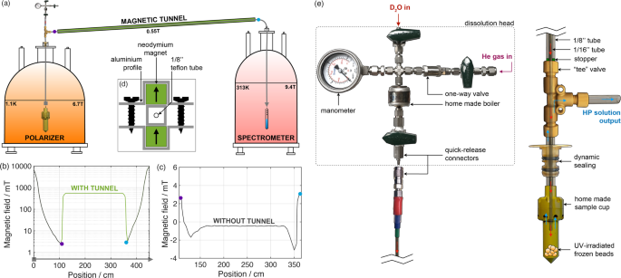
Recently, we have been developing methods to use radicals that are non-persistent. The latter, induced via UV-light irradiation (see Figure 2a) of a solution of water and pyruvic acid, recombine in biocompatible diamagnetic species at the end of the dDNP process, prior to injection.9,10 With these labile radicals, we were able to hyperpolarize water molecules up to 70% (Figure 2b), i.e. 14 times better than state-of-the-art.

Figure 2: (a) UV-irradiation of samples (b) (A) Enhanced signal and (B) non-enhanced signal.
We have the tool, now the next step will be pre-clinical implementation! Interested in reading more? Our Chemistry Communication article can be found at: https://www.nature.com/articles/s42004-020-0301-6
1. Grandin, C. B. Assessment of brain perfusion with MRI: methodology and application to acute stroke. Neuroradiology 45, 755–766 (2003).
2. van Laar, P. J., van der Grond, J. & Hendrikse, J. Brain perfusion territory imaging: Methods and clinical applications of selective arterial spin-labeling MR imaging. Radiology 246, 354–364 (2008).
3. Waters, E. A. & Wickline, S. A. Contrast agents for MRI. Basic Res Cardiol 103, 114–121 (2008).
4. Lingwood, M. D. et al. Hyperpolarized Water as an MR Imaging Contrast Agent: Feasibility of in Vivo Imaging in a Rat Model. Radiology 265, 418–425 (2012).
5. Perazella, M. A. Advanced kidney disease, gadolinium and nephrogenic systemic fibrosis: the perfect storm. Curr Opin Nephrol Hy 18, 519–525 (2009).
6. Perazella, M. A. Current Status of Gadolinium Toxicity in Patients with Kidney Disease. Clin J Am Soc Nephro 4, 461–469 (2009).
7. Ardenkjaer-Larsen, J. H. et al. Increase in signal-to-noise ratio of > 10,000 times in liquid-state NMR. Proceedings of the National Academy of Sciences of the United States of America 100, 10158–10163 (Sep 2).
8. Lipso, K. W., Hansen, E. S. S., Tougaard, R. S., Laustsen, C. & Ardenkjaer-Larsen, J. H. Dynamic coronary MR angiography in a pig model with hyperpolarized water. Magn Reson Med 80, 1165–1169 (2018).
9. Eichhorn, T. R. et al. Hyperpolarization without persistent radicals for in vivo real-time metabolic imaging. Proceedings of the National Academy of Sciences of the United States of America 110, 18064–18069 (Nov 5).
10. Capozzi, A., Cheng, T., Boero, G., Roussel, C. & Comment, A. Thermal annihilation of photo-induced radicals following dynamic nuclear polarization to produce transportable frozen hyperpolarized 13C-substrates. Nat Commun 8, 15757 (2017).
Follow the Topic
-
Communications Chemistry

An open access journal from Nature Portfolio publishing high-quality research, reviews and commentary in all areas of the chemical sciences.
Related Collections
With Collections, you can get published faster and increase your visibility.
f-block chemistry
Publishing Model: Open Access
Deadline: Feb 28, 2026
Experimental and computational methodology in structural biology
Publishing Model: Open Access
Deadline: Apr 30, 2026

Please sign in or register for FREE
If you are a registered user on Research Communities by Springer Nature, please sign in