Why Gantry Tilt Alone Is Not Enough in Head CT: Visualizing Eye Lens Dose Distribution with Monte Carlo Simulation
Published in Healthcare & Nursing and General & Internal Medicine
Why did we need to examine lens dose in head CT so carefully?
— Visualizing what had remained unseen with Monte Carlo simulation —
Head CT examinations are indispensable in clinical practice, but reducing radiation exposure to the eye lens remains a long-standing challenge because of its high radiosensitivity and its proximity to the scan range. Since the ICRP lowered the dose limit for the lens of the eye, awareness of this issue has increased even further in clinical settings.
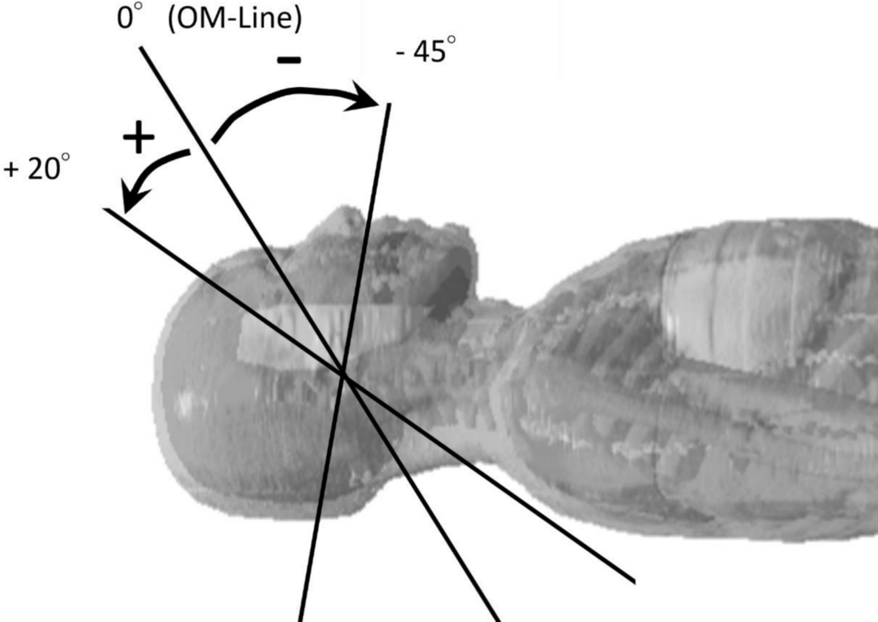
Gantry tilt has long been regarded as an effective method for reducing lens dose in head CT. However, in daily practice, we often encountered situations that raised a simple but important question: Is lens dose really reduced just by applying gantry tilt? This sense of unease became the starting point of our study.
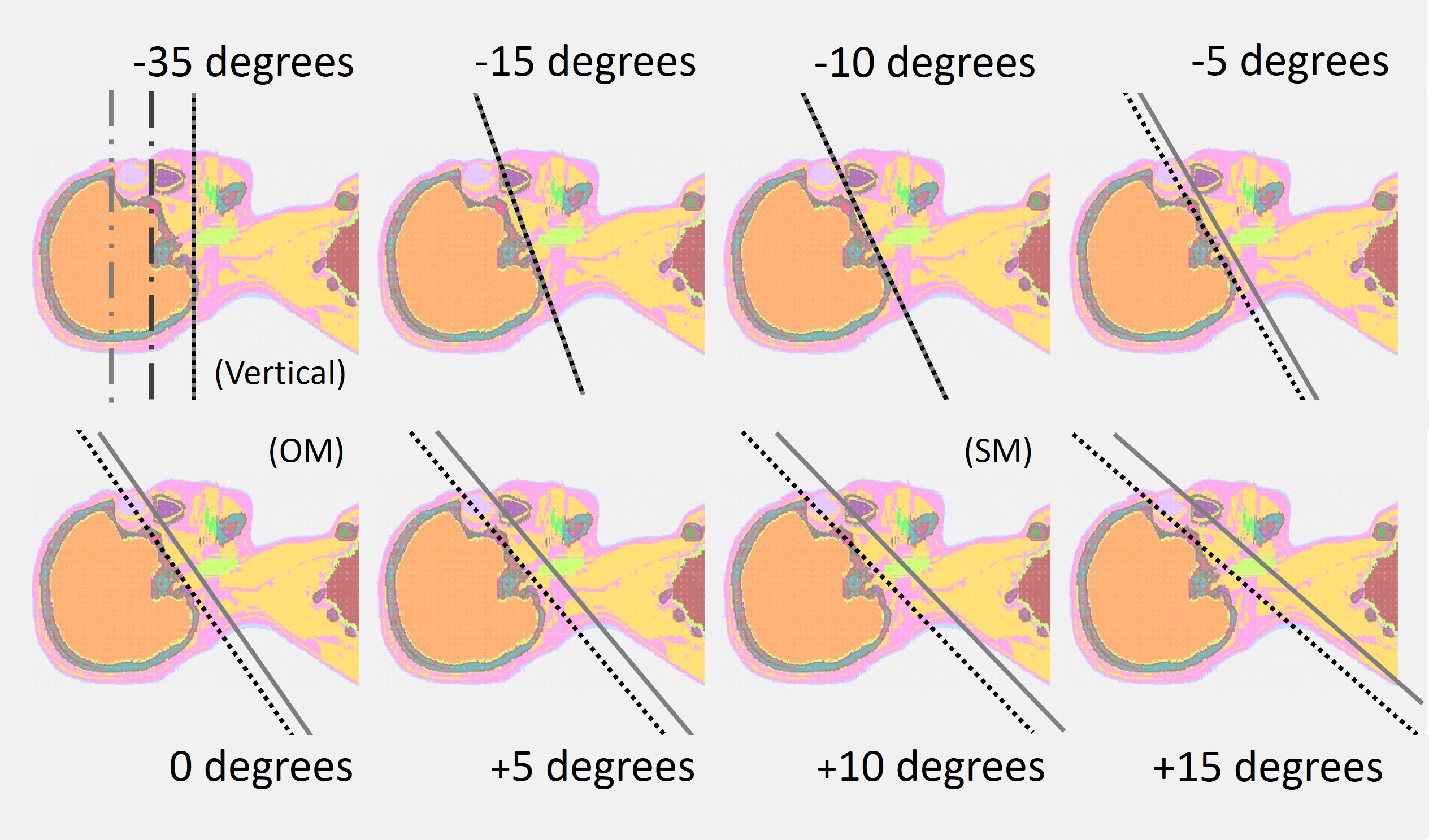
One of the difficulties in answering this question lies in the limitations of conventional dose evaluation methods. For small organs such as the eye lens, it has been difficult to assess how changes in gantry tilt angle and scan range affect the spatial dose distribution in detail. To overcome this limitation, we developed a new head CT X-ray source model using PHITS-based Monte Carlo simulation, employing a high-resolution beam with a 2-mm width in the z-direction. This allowed us to freely control gantry tilt and scan range and to visualize dose distributions with unprecedented spatial detail.
Our analysis revealed a critical finding: lens dose is not necessarily reduced by gantry tilt alone. Even with gantry tilt applied, when the scan range included the eyeballs or the lens, the lens dose did not decrease and, in some cases, even increased. This occurs because changes in beam incidence angle and scan start and end positions can shift the dose peak toward the lens region.
These results clearly indicate that effective lens dose reduction requires a combination of gantry tilt and careful restriction of the scan range to exclude the eyes and lens, rather than relying on gantry tilt alone. In addition, we also evaluated effective dose to confirm how these adjustments influence whole-body exposure, ensuring that lens dose reduction does not come at the expense of increased overall dose.
The aim of this study was not simply to report numerical values of lens dose. Rather, we sought to visualize how dose distributions change with imaging parameters and to provide a physical basis for more rational dose optimization in head CT examinations. We hope that this work will support more informed protocol design in routine clinical practice.
Follow the Topic
-
Radiological Physics and Technology
This is a multidisciplinary journal focusing on basic research and clinical applications in radiological sciences.


Please sign in or register for FREE
If you are a registered user on Research Communities by Springer Nature, please sign in