AI-supported Microscopy Enhances the Detection of Intestinal Parasitic Infections in Primary Healthcare
Published in Materials, Microbiology, and Computational Sciences
The study, published in Scientific Reports, was conducted by researchers from Karolinska Institutet (KI), the Institute for Molecular Medicine Finland (FIMM) at the University of Helsinki, Uppsala University (UU), Kinondo Kwetu Hospital, Kenya, and Muhimbili University of Health and Allied Sciences (MUHAS), Tanzania. It compares manual microscopy with two AI-based diagnostic approaches—fully autonomous AI and expert-verified AI—for detecting intestinal worm infections, so-called soil-transmitted helminths (STHs), in stool samples collected from schoolchildren in Kwale County, Kenya.
Importance of improved diagnostics
STHs, which include roundworm (Ascaris lumbricoides), whipworm (Trichuris trichiura), and hookworm, are the most prevalent neglected tropical diseases, affecting over 600 million people worldwide. The burden of STHs falls mainly on children in underserved communities, where infections can lead to malnutrition, impaired physical and mental development, and anemia. Infections are typically treated through mass-drug-administration programs targeting specific groups or entire communities. However, as global prevalence declines, test-and-treat strategies are becoming increasingly important.
To coordinate mass-drug-administration programs and individual treatment of patients, good diagnostics are needed, and today conventional microscopy of Kato-Katz-stained fecal smears is the most common method. While manual microscopy is valued for its simplicity, it has notable drawbacks: it is time-consuming, requires on-site expertise, and has low sensitivity, particularly for light-intensity infections. These limitations may be addressed through digital microscopy and AI.
The evaluated methods
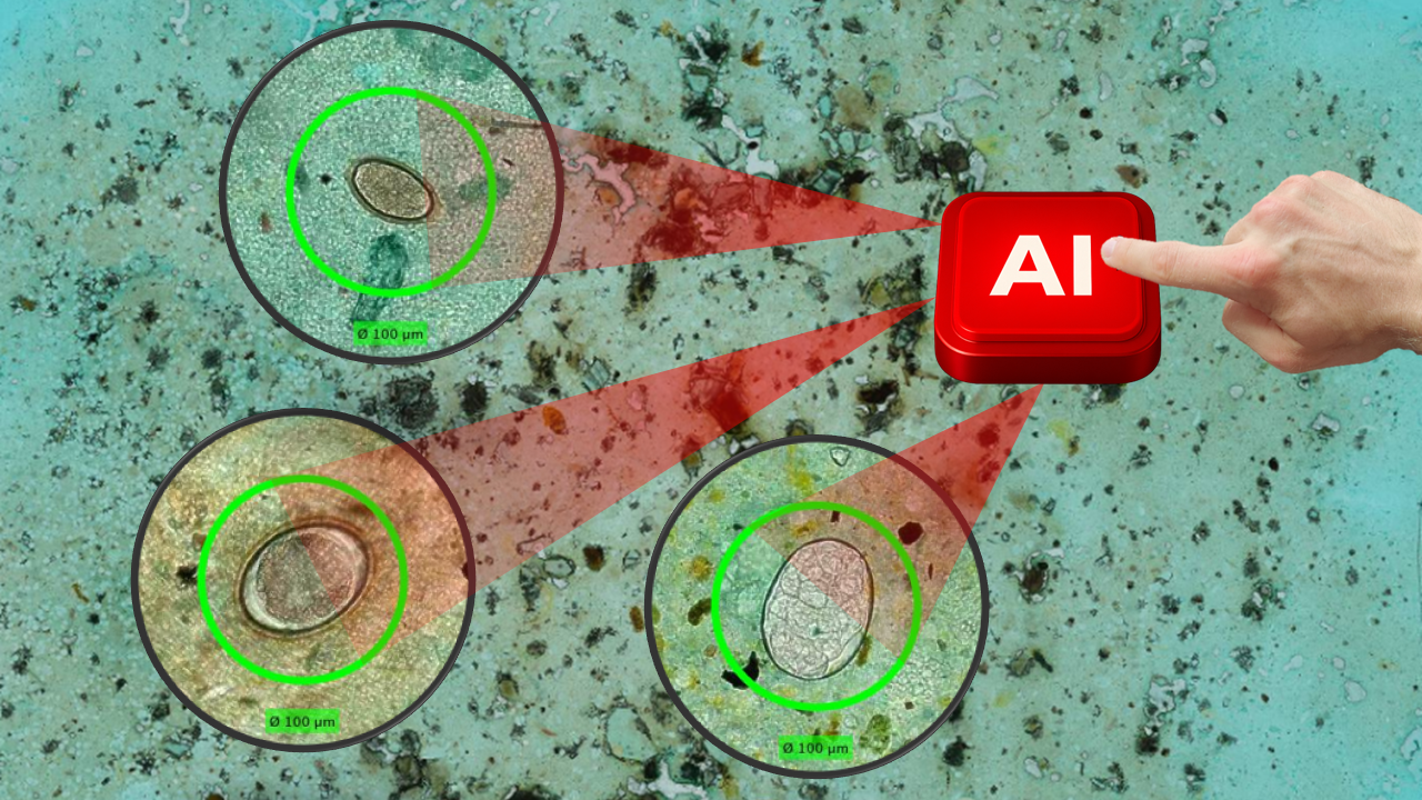
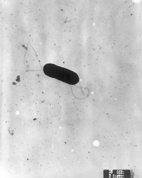
The three diagnostic methods compared in our study are illustrated in a figure from the original publication. The AI-based methods used small and portable whole-slide scanners to digitize the smears and deep-learning algorithms to analyze them. The expert-verified AI also included a verification tool where the expert could classify all the potential parasite eggs.

Findings
Among 704 valid samples, the expert-verified AI detected more infections than traditional manual microscopy, particularly for light-intensity infections that are often missed. The expert-verified AI achieved sensitivities of 92% for hookworm, 94% for T. trichiura, and 100% for A. lumbricoides, while specificity remained above 97% for all species. Manual microscopy, on the other hand, had much lower sensitivities for all species, with 78% for hookworm, 31% for T. trichiura, and 50% for A. lumbricoides.
"Our results show that the combination of AI and human expertise can surpass both manual microscopy and fully autonomous AI systems – especially in detecting light-intensity infections that might otherwise be missed," says Andreas Mårtensson, co-author and professor at UU.
Advantages of expert-verified AI
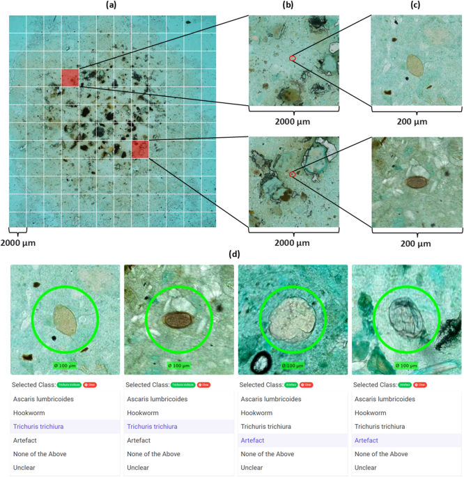
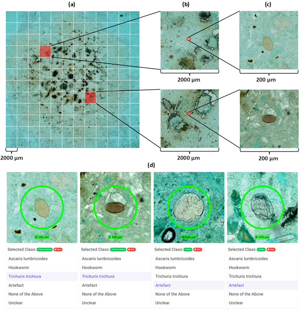
The expert-verified AI system allows local experts to confirm AI findings in less than one minute, drastically reducing workload while increasing accuracy. "The fact that the expert-verified AI had the highest sensitivity for all species shows how AI can help find the needle (parasite egg) in the haystack, enhancing human capabilities and diagnostics,” says Dr. Joar von Bahr, PhD student affiliated with KI, FIMM, and UU and first author of the article. “I think our figure from the published manuscript illustrates what a tedious task it is for experts to identify parasite eggs if only a few are present in a smear. On the other hand, with the help of the AI, it becomes almost impossible to miss an egg if it is presented to the expert," says von Bahr. The figure from the article shows (parts a-c) how the experts need to analyze more than 100 fields-of-view to find one of the two present parasite eggs, but in the expert-verified AI (part d), only four objects are presented, two of which were parasite eggs.
Implications
The high sensitivity and specificity of the expert-verified AI in this study indicate it may fulfill the diagnostic accuracy of an ideal diagnostic method according to the WHO's target product profile for STH diagnostics. “Our work has shown how cutting-edge technology can be brought directly into our rural hospital setting to improve patient care. By using AI and digital microscopy, screening for STH infections can be made more efficient and accurate, which is critical for children’s health in our community. Our research not only strengthens the local diagnostic capacity but also has significant global relevance,” remarks Harrison Kaingu, CEO, Kinondo Kwetu Hospital, Kwale County, Kenya. “Our method could provide accurate, fast, and scalable diagnostics at the point of care, particularly important as global STH prevalence declines and more sensitive methods are required for disease monitoring," says Dr. Nina Linder, senior co-author and guest professor at UU.
The findings mark a significant step forward in using AI to address diagnostic needs for neglected tropical diseases, where our low-infrastructure expert-verified AI-based digital microscopy showed higher sensitivity for all STH species compared to manual microscopy. Additionally, it can be run on-site in a primary health care laboratory with a total sample analysis time of 15 minutes and less than one minute of hands-on work by the expert. “This research shows the potential of combining portable imaging with AI to overcome long-standing diagnostic challenges in global health,” says Dr. Johan Lundin, professor and senior co-author from KI and FIMM.
The study was carried out in collaboration with partners in Kenya, Sweden, Finland, and Tanzania, and was supported by the Erling-Persson Foundation, the Swedish Research Council, and several private foundations in Finland.
Contact:
Joar von Bahr
Department of Global Public Health
Karolinska Institutet
joar.von.bahr@ki.se
Images 1 and 2: from the original article
-
I have a medical degree from Karolinska Institutet and am currently a PhD student at Karolinska Institutet and the University of Helsinki under the supervision of Professors Nina Linder and Johan Lundin.
-
The focus of my PhD is developing artificial intelligence supported image-based diagnostics for low-resource settings. Primarily for cervical cancer screening and soil-transmitted helminth diagnosis.
-
I am interested in the integration of medicine and technical advancements to improve diagnosis, especially with a focus on artificial intelligence.
- https://ki.se/en/people/joar-von-bahr#about-me
Follow the Topic
-
Scientific Reports
An open access journal publishing original research from across all areas of the natural sciences, psychology, medicine and engineering.

Your space to connect: The Polarised light Hub
A new Communities’ space to connect, collaborate, and explore research on Light-Matter Interaction, Optics and Photonics, Quantum Imaging and Sensing, Microscopy, and Spectroscopy!
Continue reading announcementRelated Collections
With Collections, you can get published faster and increase your visibility.
Phytochemicals and health
Publishing Model: Open Access
Deadline: Jul 28, 2026
Health Policy and Systems Research
Publishing Model: Hybrid
Deadline: Jul 14, 2026
Please sign in or register for FREE
If you are a registered user on Research Communities by Springer Nature, please sign in