Another giant emerges from the heart of South America
Published in Microbiology

By Matheus Rodrigues, Victoria Queiroz, Frank Aylward and Jonatas Abrahao, on behalf of all co-authors.
Brazil is a country of astonishing biodiversity, and few places capture this more vividly than the Pantanal. Recognized as one of the world’s biodiversity hotspots, this unique wetland is under constant threat—from climate change to human activities. Years ago, it was in this setting that we discovered Tupanvirus, one of the most extraordinary giant viruses described. That experience opened our eyes to the hidden microbial richness of the Pantanal and made us eager to return, curious about what other invisible worlds might be hiding.
This time, we joined the NAVIO Project (Expanded Navigation for Intensive and Optimized Surveillance), led by Prof. Luiz Alcantara (FIOCRUZ). Developed in partnership with the Brazilian Navy, NAVIO not only brings together researchers from many disciplines to study how microorganisms circulate in aquatic environments and their implications for ecosystems and public health, but also provides essential healthcare services to riverine communities along its route. During the expedition along the Paraguay River, we collected water, soil, sediment, and sewage samples, always diving into the unknown. We knew the region was full of life, but we couldn’t predict what we would find in the lab.

Matheus Rodrigues (co-first author of the paper) collecting water from the Paraguay River.
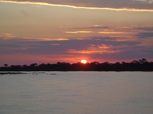
Sunset over the Paraguay River, Pantanal biome, Brazil.
Back in the lab, we began screening for giant viruses in Vermamoeba vermiformis. The process was tricky—fungal and bacterial contamination was a constant challenge, and we knew this amoeba only allows very few viruses to replicate. After many frustrating attempts, we finally observed a cytopathic effect in one of the samples. Excited, we expanded the virus and also tested it in Acanthamoeba castellanii. The effect repeated. At first, we thought it might be Tupanvirus—the only known giant virus able to infect two different amoeba genera. But something didn’t fit: the typical “cell clumps” caused by Tupanvirus were absent.
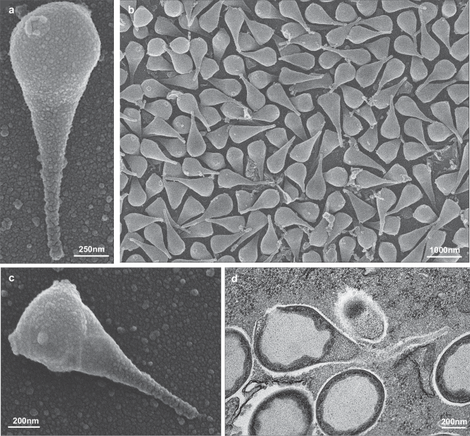
That’s when we turned to electron microscopy for answers. Scanning electron microscopy revealed something entirely unexpected: drop-shaped particles, with a flexible tail and two ostioles at the top. We were amazed. Hypotheses piled up, but only transmission electron microscopy confirmed what we suspected—a completely new virus. Cross-sections revealed a uniquely enveloped capsid, a truly novel architecture. More questions arose. How critical was the envelope for infection? To explore this, we used immunofluorescence microscopy and treated the particles with detergents. Each experiment revealed new surprises and deepened our sense that this virus was unlike anything we had seen before.
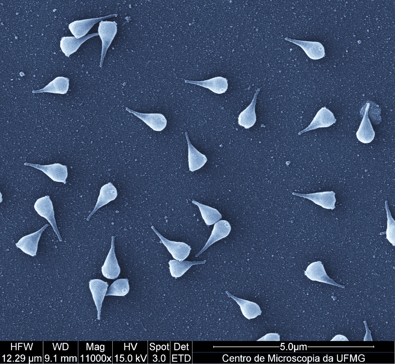
Scanning electron microscopy of Naiavirus particles observed during the first microscopy session at the end of 2024. Centro de Microscopia, UFMG.
Morphology alone, however, wasn’t enough. We moved on to genome sequencing, an enormous effort. We achieved very high coverage, allowing us to assemble a complete, circular genome of ~922,000 base pairs. The genome contains ~867 genes, many of which are ORFans, with no known homologs. Our analyses confirmed that this virus represents a new lineage within the Pimascovirales, distinct from any previously described giant virus.
In parallel, we performed proteomic analyses of the viral particle, mapping all proteins present in the capsid and particle. We also conducted a large-scale search across over 16,000 metagenomic datasets from soils, frozen environments, lake waters, and more. We found sequences related to Naiavirus across the globe, particularly in cold regions. This showed that viruses related to Naiavirus are globally distributed—but the one we isolated was the first fully characterized in the lab, with its unique morphology, ability to infect amoebas, remarkable genome, and protein composition.
Naiavirus fascinates not just for its distinct shape—flexible tail, envelope, and ostioles—but also for its rich, novel genome and proteome. It reminds us that even in well-studied ecosystems, invisible worlds await discovery. Every expedition, every sample, and every collaborative effort has the potential to reveal these hidden treasures. Naiavirus exemplifies how curiosity, teamwork, and technology together can uncover the unknown.
Follow the Topic
-
Nature Communications
An open access, multidisciplinary journal dedicated to publishing high-quality research in all areas of the biological, health, physical, chemical and Earth sciences.

Related Collections
With Collections, you can get published faster and increase your visibility.
Women's Health
Publishing Model: Hybrid
Deadline: Ongoing
Tumor Microenvironment Crosstalk and Therapeutic Implications
Publishing Model: Hybrid
Deadline: Nov 02, 2026

Please sign in or register for FREE
If you are a registered user on Research Communities by Springer Nature, please sign in