Aptahistochemistry (AHC): The Next-Generation Molecular Disruptor in Tissue Diagnostics
Published in General & Internal Medicine and Immunology
Introduction
For decades, Immunohistochemistry (IHC) has served as the gold standard for detecting protein expression in tissues a cornerstone technique in diagnostic pathology, oncology, and biomedical research. However, despite its reliability, IHC has limitations: antibody variability, cross-reactivity, high production costs, and the need for cold-chain storage.
Enter Aptahistochemistry (AHC) a revolutionary alternative that employs synthetic DNA or RNA aptamers instead of antibodies. This innovation promises greater precision, reproducibility, and affordability while integrating seamlessly with digital and AI-assisted histopathology workflows.
What Is Aptahistochemistry (AHC)?
Aptahistochemistry is an advanced molecular technique that utilizes aptamers short, single-stranded oligonucleotides (DNA or RNA) to bind specifically to target molecules, much like antibodies do in IHC. These aptamers are identified through a process called SELEX (Systematic Evolution of Ligands by Exponential Enrichment), which selects sequences with the highest binding affinity to a given antigen.
Key Features of Aptamers
-
Synthetic origin: Chemically synthesized without animal hosts or hybridoma systems.
-
Customizability: Their sequences can be modified to enhance binding strength, stability, and fluorescence labeling.
-
Stability: Aptamers withstand higher temperatures, pH changes, and long storage times.
-
Reversibility: They can be denatured and reused under certain conditions.
How Does AHC Work?
The workflow of AHC closely parallels that of IHC, but with aptamers replacing antibodies.
-
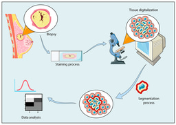
Tissue Preparation:
Formalin-fixed paraffin-embedded (FFPE) or frozen tissue sections are deparaffinized and rehydrated. -
Antigen Retrieval:
Heat-induced or enzymatic retrieval may be applied to expose epitopes or aptamer-binding sites. -
Aptamer Incubation:
Fluorophore- or enzyme-labeled aptamers are incubated with the tissue section. These aptamers bind their target proteins with nanomolar affinity. -
Signal Detection:
Bound aptamers are visualized via chromogenic substrates (e.g., DAB) or fluorescent imaging systems, just like in IHC or IF. -
Imaging & Analysis:
Modern AHC setups often employ computer-assisted imaging and AI-based pattern recognition, enhancing quantitative and reproducible analysis of molecular expression.
Advantages of AHC Over Conventional IHC
| Feature | IHC (Antibody-based) | AHC (Aptamer-based) |
|---|---|---|
| Reagent Type | Animal-derived antibodies | Synthetic DNA/RNA aptamers |
| Production | Expensive, time-consuming | Rapid and cost-effective chemical synthesis |
| Batch Variability | High (lot-to-lot variation) | Minimal (sequence-defined) |
| Specificity | Sometimes cross-reactive | High due to engineered binding sites |
| Storage | Requires cold chain | Stable at room temperature |
| Signal Detection | Enzyme/fluorophore-labeled antibodies | Fluorophore-labeled aptamers or nano-tags |
| Automation Potential | Moderate | High (AI-integrated digital imaging) |
| Cost | Relatively expensive | Significantly cheaper (after initial SELEX setup) |
Why AHC Could Replace Conventional IHC
-
Precision & Reproducibility:
Aptamers are sequence-defined molecules, ensuring identical performance across labs and time. -
Lower Costs:
Chemical synthesis eliminates animal facilities and hybridoma maintenance, drastically reducing production costs. -
Enhanced Stability:
Aptamers remain stable even under harsh storage or assay conditions, ideal for resource-limited settings. -
AI Integration:
AHC workflows are inherently compatible with digital pathology aptamer labeling can produce uniform, quantifiable signals, enhancing computational image analysis. -
Ethical & Sustainable:
No animals are used for aptamer generation, aligning with the principles of 3Rs (Replacement, Reduction, Refinement) in biomedical research.
Future Prospects: Toward Smart Histopathology
Aptahistochemistry represents a convergence of molecular pathology, nanotechnology, and AI.
Future directions include:
-
Multiplexed AHC panels for simultaneous detection of multiple biomarkers.
-
Integration with NGS and proteomics for molecular diagnostics.
-
Clinical translation in oncology, infectious disease pathology, and precision medicine.
As laboratories continue to digitize and move toward morpho-molecular diagnostics, AHC has the potential to supersede conventional IHC offering faster, cheaper, and more reproducible results, with molecular-level insight and computational scalability.


Please sign in or register for FREE
If you are a registered user on Research Communities by Springer Nature, please sign in