Capturing the Edge of Polymorphisms: A Reference-Free Variant Detection Method
Published in Cancer, Protocols & Methods, and Genetics & Genomics

Next-generation sequencing (NGS) has revolutionized genome analysis by enabling rapid and cost-effective acquisition of whole-genome data. However, the output consists of fragmented reads—typically ranging from 100 bases to several tens of kilobases—rather than continuous genome sequences. Variant detection is commonly performed by mapping these fragments to a reference genome, but this approach becomes infeasible when no reference is available.
A method was developed to directly compare NGS reads from two samples, allowing for the detection of polymorphisms without constructing a reference genome. This enables direct investigation of genotype–phenotype correlations. The challenge resembles identifying mismatched pieces from two jigsaw puzzles cut from slightly different images—an endeavor long considered impractical due to the sheer volume of data.
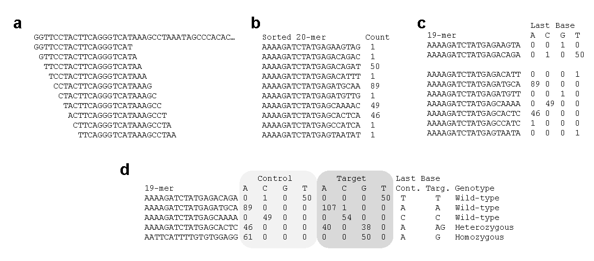
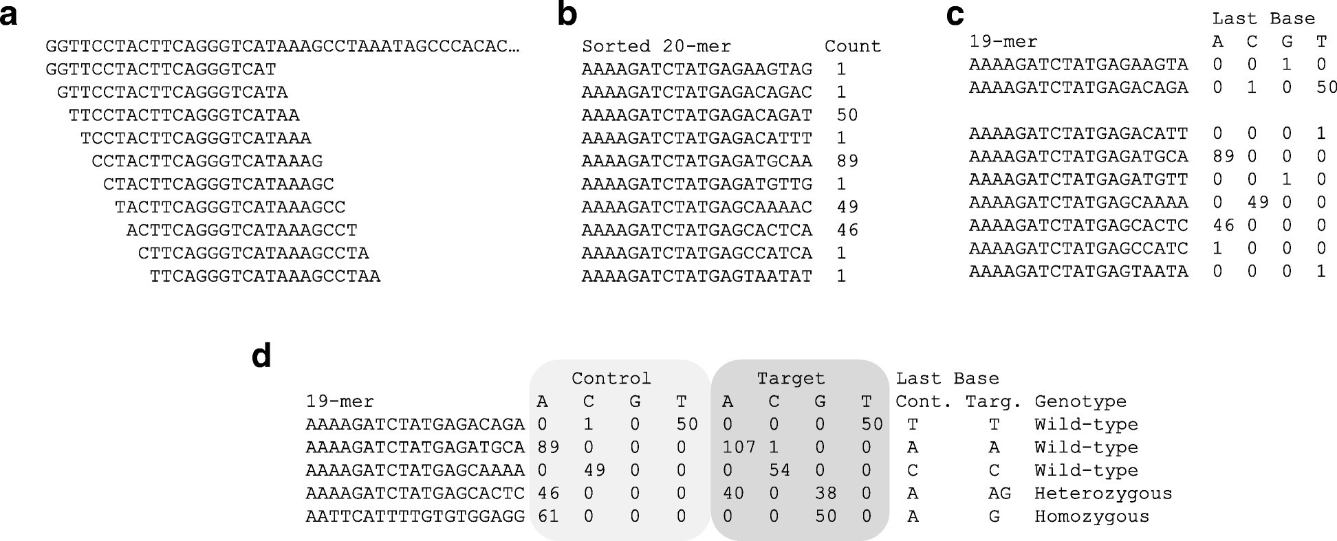
To simplify comparisons, each read is segmented into 20-base sequences (20-mers) by sliding one base at a time. For example:
- Sample A: AAATGGTACATTTATATTAT
- Sample B: AAATGGTACATTTATATTAC
A difference in the final base indicates a polymorphism. This approach detects not only single nucleotide polymorphisms (SNPs), but also edge of structural variants such as insertions, deletions, inversions, and translocations. While the exact nature of the mutation may not be immediately clear, its presence is evident.
The extracted 20-mers are sorted and grouped by identical sequences, with counts displayed (Fig. 1a and b). These are then split into the first 19 bases and the final base, and the counts are aggregated per nucleotide (Fig. 1b and c). For instance, if two sequences share the same first 19 bases but differ at the final base (e.g., one ends in C and the other in T), the output will show a single row with counts for C and T respectively (Fig. 1c).
Control and Target datasets are then compared using the same 19-mer sequences. Most sequences will have matching final bases, but polymorphic sites will show differing counts (Fig. 1d). These counts correspond to read depth, and a count of 1 is typically considered a sequencing error. If a reference genome is available, the genomic location of the 19-mer can be used to pinpoint the polymorphic edge. If not, the 19-mer itself serves as a unique identifier for the polymorphism.
Although NGS generates massive data volumes, and extracting 20-mers by sliding one base increases the data size twentyfold, a method was established—after years of trial and error—that enables analysis on a single computer.
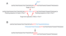
In April, a bidirectional alignment method for detecting polymorphic edges was introduced. The current method also targets edge detection, and both approaches are presented in a single paper for comparative evaluation.
By grouping individuals based on the presence or absence of a trait and obtaining sufficient sequencing data, it becomes possible to identify trait-linked polymorphisms without a reference genome.
This reference-free variant detection method is particularly effective for non-model organisms and rapid screening scenarios. For those interested, the following publication and resources provide further details:
Reference
Miyao, A., Kiyomiya, J.S., Iida, K. et al. Polymorphic edge detection (PED): two efficient methods of polymorphism detection from next-generation sequencing data. BMC Bioinformatics 20, 362 (2019). https://doi.org/10.1186/s12859-019-2955-6
GitHub: https://github.com/akiomiyao/ped
Follow the Topic
-
BMC Bioinformatics
This is an open access, peer-reviewed journal that considers articles describing novel computational algorithms and software, models and tools, including statistical methods, machine learning and artificial intelligence, as well as systems biology.

Related Collections
With Collections, you can get published faster and increase your visibility.
Computational methods in paleobiology
BMC Bioinformatics is welcoming submissions to our Collection on Computational methods in paleobiology.
The interdisciplinary field of paleobiology enables researchers to reconstruct evolutionary histories, model population dynamics, and explore the multifactorial influences that shaped life in the past. Paleobiology includes, but is not limited to, macrofossils, microfossils, and ancient proteins, DNA and RNA from both fossils and environmental sources. For instance, with the rapid growth of paleogenomic datasets, novel computational approaches are essential for extracting insights from this fragmented ancient data.
This Collection welcomes submissions on the development of new computational and/or statistical approaches for paleobiology. We encourage contributions that highlight innovative methods for analyzing ancient DNA, modeling evolutionary processes, integrating heterogeneous datasets, and visualizing temporal and spatial patterns.
All manuscripts submitted to this journal, including those submitted to collections and special issues, are assessed in line with our editorial policies and the journal’s peer-review process. Reviewers and editors are required to declare competing interests and can be excluded from the peer review process if a competing interest exists.
Publishing Model: Open Access
Deadline: Jan 23, 2027
Cell tracking
BMC Bioinformatics is welcoming submissions to our Collection on Cell Tracking.
Cell tracking is a technique used to monitor and analyze the movement and behavior of cells over time, allowing the study of cellular behaviors, dynamics, and interactions within various biological contexts. Advanced bioinformatics tools play a vital role in analyzing cell tracking data. They help identify cell movement patterns and understand their biological implications. These tools are particularly relevant when processing large datasets and when investigating cell cycles.
This Collection welcomes submissions on the development of new computational and/or statistical approaches for cell tracking. We encourage contributions detailing methods for detecting and characterizing cell movements to better understand cell migration and behavior.
All manuscripts submitted to this journal, including those submitted to collections and special issues, are assessed in line with our editorial policies and the journal’s peer-review process. Reviewers and editors are required to declare competing interests and can be excluded from the peer review process if a competing interest exists.
Publishing Model: Open Access
Deadline: Sep 23, 2026


Please sign in or register for FREE
If you are a registered user on Research Communities by Springer Nature, please sign in