Detecting Transposable Element Insertions from NGS Data
Published in Genetics & Genomics and Plant Science

Transposable elements, often referred to as “jumping genes,” have the remarkable ability to insert copies of their sequences into different locations within the genome.
With next-generation sequencing (NGS), obtaining the complete genomic sequence of an organism has become routine. This means we should also be able to track how transposable elements move within the genome using these sequence data.
However, NGS produces millions to billions of short reads, each only a few hundred bases long—like an enormous jigsaw puzzle. Reconstructing continuous chromosome sequences from these fragments requires extensive computation.
The presence of transposable elements complicates this process. When elements longer than the read length are scattered throughout the genome, assembly algorithms often generate multiple possible solutions, making it difficult to determine the correct one.
Long-read sequencing technologies can resolve this issue, but with short-read data alone, it remains challenging. Moreover, because identical copies of the target transposon are dispersed across the genome, simple similarity searches cannot reliably reveal where and how the element has moved.
After days of thinking about this problem, I realized that the target site duplication (TSD) created during transposition could serve as a key indicator.
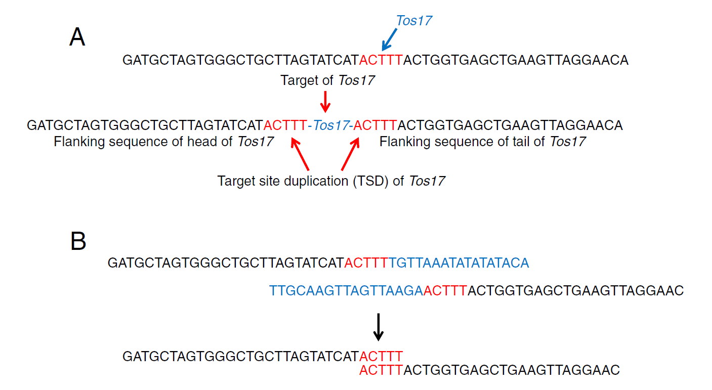
Figure 1A shows the TSD associated with the Tos17 retrotransposon, which was my focus at the time. When Tos17 inserts, a 5-base duplication appears flanking its upstream and downstream ends.
By extracting short reads that contain the 5′ and 3′ ends of Tos17 and removing the Tos17 sequence itself, I collected the adjacent sequences (Figure 1B). When two sequences share the same 5-base motif at their ends, they form a pair—indicating the insertion site of Tos17.
If a reference genome is available, these paired sequences can be mapped directly to identify insertion sites. Even without a reference, the paired sequences themselves mark the location of Tos17 and can be used as markers.
The approach is simple: search short reads for transposon termini and pair the adjacent sequences. This can be implemented with a lightweight program.
I developed a prototype in Perl with fewer than 50 lines of code. Because it relies only on basic searches and pairing a small number of sequences, I believe this is one of the smallest and fastest programs for analyzing NGS data.
Feel free to download and try it:
https://github.com/akiomiyao/tif
Reference
Nakagome, M., Solovieva, E., Takahashi, A. et al. Transposon Insertion Finder (TIF): a novel program for detection of de novo transpositions of transposable elements. BMC Bioinformatics 15, 71 (2014). https://doi.org/10.1186/1471-2105-15-71

Follow the Topic
-
BMC Bioinformatics
This is an open access, peer-reviewed journal that considers articles describing novel computational algorithms and software, models and tools, including statistical methods, machine learning and artificial intelligence, as well as systems biology.

Related Collections
With Collections, you can get published faster and increase your visibility.
Extracellular vesicle research
BMC Bioinformatics is welcoming submissions to our Collection on Extracellular vesicles research.
BMC Bioinformatics is welcoming submissions to our Collection on Extracellular vesicles research. Extracellular vesicles (EVs) are are small lipid bilayer-delimited particles released by cells that play crucial roles in intercellular communication and various physiological processes. The study of EVs has gained significant attention due to their potential as biomarkers for disease diagnosis, therapeutic targets and drug delivery systems. Advanced bioinformatics tools are essential for analyzing EV data, identifying EV-associated molecules, and understanding their biological functions.
This Collection welcomes submissions on the development of new computational and/or statistical approaches for the study of extracellular vesicles. We encourage contributions that highlight innovative methods for detecting and characterizing EVs and elucidating the molecular mechanisms underlying EV biogenesis and function.
All manuscripts submitted to this journal, including those submitted to collections and special issues, are assessed in line with our editorial policies and the journal’s peer-review process. Reviewers and editors are required to declare competing interests and can be excluded from the peer review process if a competing interest exists.
Publishing Model: Open Access
Deadline: Dec 30, 2026
Computational methods in paleobiology
BMC Bioinformatics is welcoming submissions to our Collection on Computational methods in paleobiology.
The interdisciplinary field of paleobiology enables researchers to reconstruct evolutionary histories, model population dynamics, and explore the multifactorial influences that shaped life in the past. Paleobiology includes, but is not limited to, macrofossils, microfossils, and ancient proteins, DNA and RNA from both fossils and environmental sources. For instance, with the rapid growth of paleogenomic datasets, novel computational approaches are essential for extracting insights from this fragmented ancient data.
This Collection welcomes submissions on the development of new computational and/or statistical approaches for paleobiology. We encourage contributions that highlight innovative methods for analyzing ancient DNA, modeling evolutionary processes, integrating heterogeneous datasets, and visualizing temporal and spatial patterns.
All manuscripts submitted to this journal, including those submitted to collections and special issues, are assessed in line with our editorial policies and the journal’s peer-review process. Reviewers and editors are required to declare competing interests and can be excluded from the peer review process if a competing interest exists.
Publishing Model: Open Access
Deadline: Jan 23, 2027