Generative AI: A Game-Changer in Age-Related Macular Degeneration Screening by Achieving ‘Noninvasive’ Indocyanine Green Angiography
Published in Bioengineering & Biotechnology, Protocols & Methods, and General & Internal Medicine

Drawbacks and Challenges of Applying Unimodal Imaging for AMD Detection
Age-related macular degeneration (AMD) is the leading cause of central vision loss in the aging population. Color fundus photography (CF) is widely applied for AMD screening due to its simplicity and low cost, particularly in source limited area. But CF images have limitations in detecting and distinguishing certain lesions, because of the unstable image quality and common characteristics shared by several chorioretinal diseases on CF images. Indocyanine green angiography (ICGA) is another well-established fundus imaging technique for screening chorioretinal conditions, owning its unique advantages in visualizing deeper choroidal vasculature and lesions behind retinal pigment epithelium. However, ICGA is an invasive imaging modality with potential adverse reactions. Besides, the complex operating procedures impede its widespread implementation in clinical settings.
Leverage the Power of GANs: Providing ‘Noninvasive’ ICGA to Enable Multimodal AMD Screening
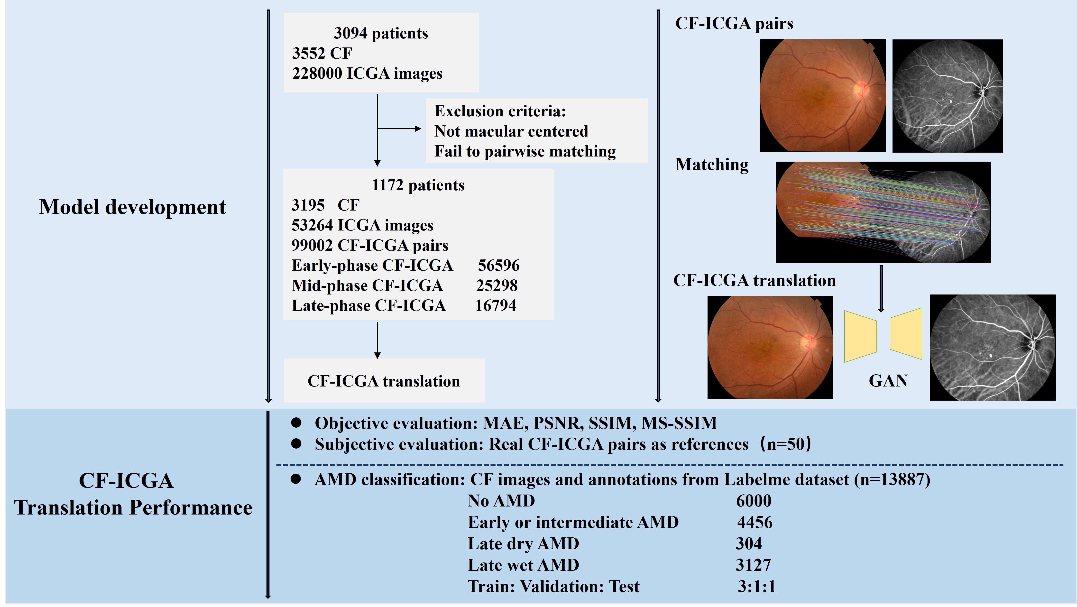
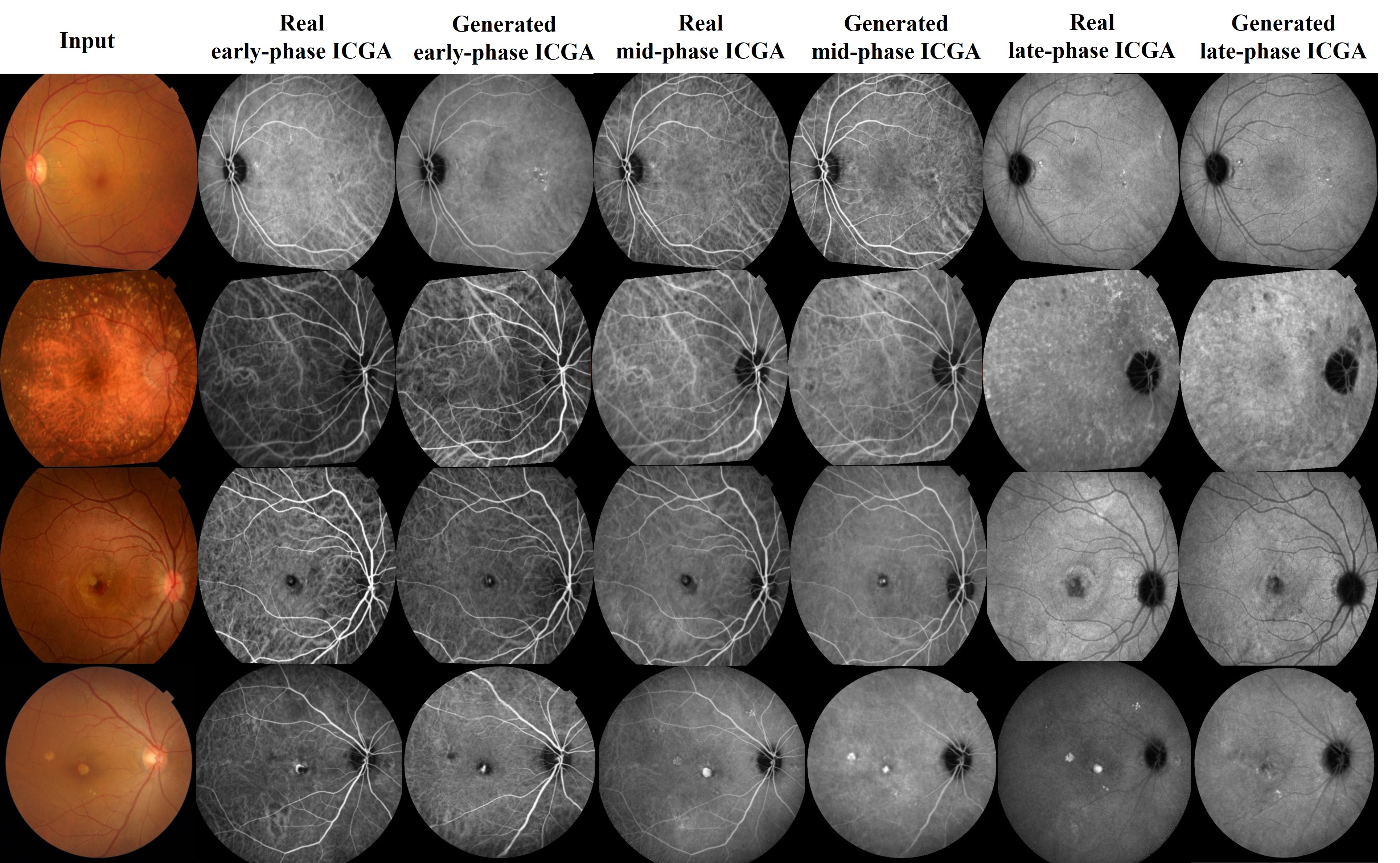
To harness the benefits of different imaging modalities and achieve efficient multimodal AMD screening, we innovatively developed a cross-modality translation model capable of synthesizing realistic ICGA images from non-invasive and easily accessible CF images using generative adversarial networks (GANs) (Figure 1). The algorithm showed high authenticity in generating anatomical structures and pathological lesions in both internal and external datasets (Figure 2). Additionally, the integration of translated ICGA images with real CF images significantly improves the accuracy of AMD screening and effectively reduce classification errors in external validation (Table 1).

Figure 1. Flow chart of the study. GAN=generative adversarial networks, CF=color fundus photography, ICGA=indocyanine green angiography, AMD=age-related macular degeneration, MAE=mean absolute error, PSNR= peak signal-to-noise ratio, SSIM= structural similarity measures, MS-SSIM=multi-scale structural similarity measures.

Figure 2. Examples of real and translated indocyanine green angiography (ICGA). 1st row, early dry AMD, 2nd row, intermediate dry AMD, 3rd row, wet AMD, 4th row, wet AMD. 1-3 rows: internal test set, 4th row: external test set.
|
|
F1-score |
Sensitivity |
Specificity |
Accuracy |
AUC |
P value△ |
|
CF |
0.8386 |
0.8368 |
0.9323 |
0.8368 |
0.9312 |
|
|
CF+early |
0.8601 |
0.8598 |
0.9428 |
0.8598 |
0.9407 |
0.4400 |
|
CF+early+mid |
0.8854 |
0.8850 |
0.9466 |
0.8850 |
0.9632 |
<0.0001* |
|
CF+early+mid+late |
0.8875 |
0.8872 |
0.9474 |
0.8872 |
0.9688 |
<0.0001* |
Conclusion and future work
Our study pioneeringly established the feasibility of generating ICGA using CF, and introduced a cross-modality approach to augment data for AMD-related deep learning research. Furthermore, our findings highlight the potential of the CF-to-ICGA model as a valuable approach for evaluating multiple chorioretinal diseases accurately and non-invasively. Further prospective trials are required to translate this research discovery into clinical benefit in real-world practice.
Follow the Topic
-
npj Digital Medicine

An online open-access journal dedicated to publishing research in all aspects of digital medicine, including the clinical application and implementation of digital and mobile technologies, virtual healthcare, and novel applications of artificial intelligence and informatics.
Your space to connect: The Macular degeneration Hub
A new Communities’ space to connect, collaborate, and explore research on Ophthalmology and Eye Diseases!
Continue reading announcementRelated Collections
With Collections, you can get published faster and increase your visibility.
Digital Health Equity and Access
Publishing Model: Open Access
Deadline: Mar 03, 2026
Evaluating the Real-World Clinical Performance of AI
Publishing Model: Open Access
Deadline: Jun 03, 2026


Please sign in or register for FREE
If you are a registered user on Research Communities by Springer Nature, please sign in