Hippo signaling pathway in cancer cells induces the formation of specific CAF subset to modulate immune activity in the tumor microenvironment
Published in Cancer
Nowadays, cancer immunotherapy has become a significant focus in cancer treatment. With the aim to utilize activated immune cells to kill cancer cells. However, small patients respond to current immunotherapies. The problem is that cancer cells adapt themselves to survive by evading those active immune cells.
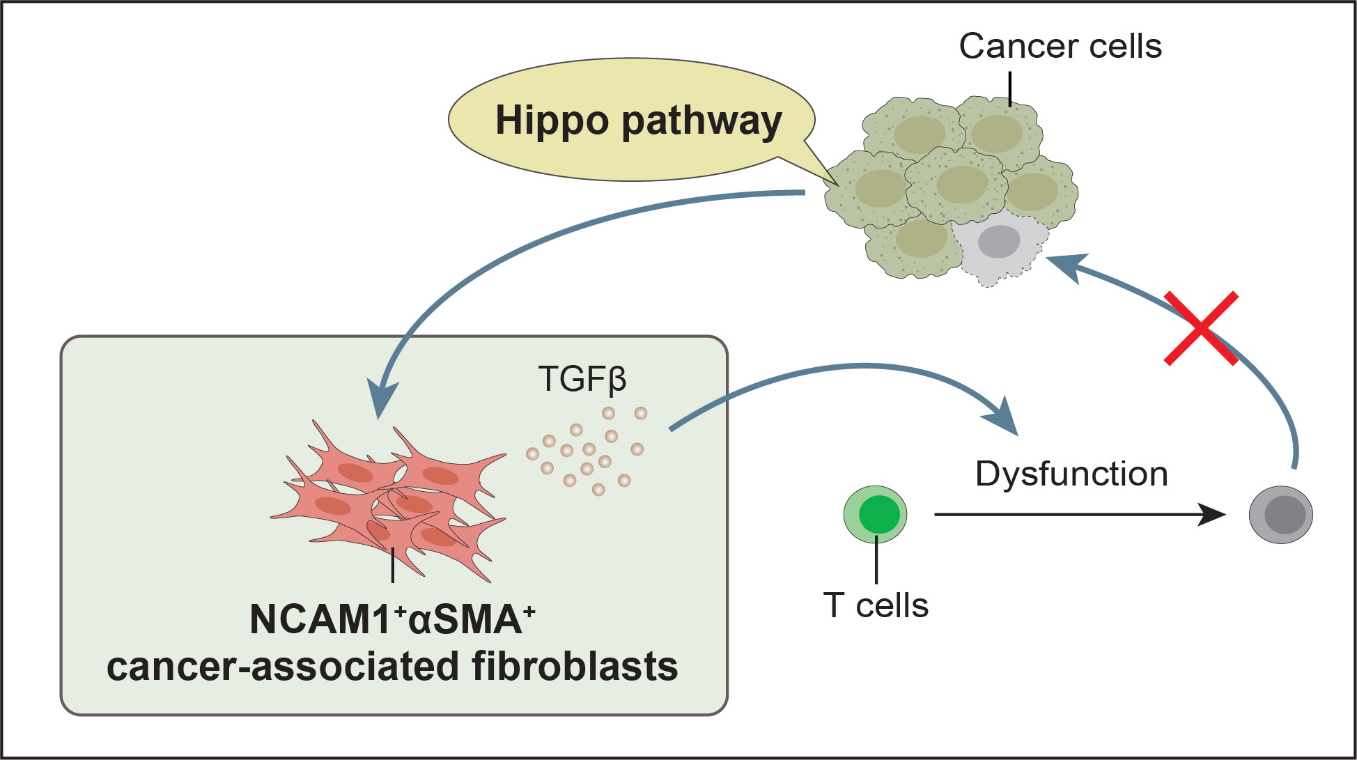
We previously published a paper showing that Hippo pathway kinases, large tumor suppressor 1 and 2 (LATS1/2), in cancer cells can suppress the initial activity of immune cells against cancer. LATS1/2 kinases attenuate the generation of cytotoxic T cells, which have the ability to kill cancer cells. This event occurs in the lymphoid organs during the early stages of tumor formation—LATS1/2 double knockout (dKO) in cancer cells promotes the production of cytotoxic T cells and suppress tumor growth by day 12 after tumor formation. Interestingly, we found that LATS1/2 dKO not only suppressed tumor growth at early stage (12 days), but it also suppressed growth throughout tumor progression (over a month). Indeed, we found some activated T cells infiltrating LATS1/2 wild-type (WT) tumors at the late stage of tumor progression. Thus, even though some activated T cells can enter tumor site, they cannot kill cancer cells in LATS1/2 WT tumors – Why?
Cancer cells reside within their environments, known as the tumor microenvironment (TME). TME is composed of cancer cells and their environments with various cell types, including immune cells, endothelial cells, pericytes, and cancer-associated fibroblasts (CAFs) which are known to promote their fitness. This made us curious that the interactions of those environmental cells and cancer cells enable cancer cells to escape immune attacks. Since CAFs have a long-standing reputation for manipulating immune activity in TME – we asked: can LATS1/2 in cancer cells regulate CAFs and whether this event suppresses immune activity in TME?
To answer this, we first investigated whether LATS1/2 dKO in cancer cells alters CAF compositions in the TME. To this end, we performed single-cell RNA sequencing (scRNA-seq) analysis to characterize CAF subpopulations in LATS1/2 dKO tumors compared to WT tumors. We used a syngeneic mouse breast tumor model (BALB/c mice with 4T1 breast cancer) due to its abundance of CAFs. As a result, scRNA-seq revealed the CAF heterogeneity in the TME of the mouse breast tumors, with 6 distinct CAF subsets. We found that NCAM1+αSMA+ CAF subpopulation was reduced in LATS1/2 dKO tumors, suggesting that LATS1/2 in cancer cells induce NCAM1+αSMA+ CAF formation. We then asked: what is the biological function of NCAM1+αSMA+ CAFs? Can they modulate immune activity in the TME?
To answer this, we performed scRNA-seq analysis to compare transcriptional profiles of our NCAM1+αSMA+ CAFs to other well-studied CAF subsets in pancreatic ductal adenocarcinoma; myofibroblastic CAFs (myCAFs), inflammatory CAFs (iCAFs), antigen-presenting CAFs (apCAFs), and leucine-rich repeat containing 15 (LRRC15)+ CAFs. We found that NCAM1+αSMA+ CAFs showed a transcriptional similarity to myCAFs and LRRC15+ CAFs, both of which possess myofibroblast-like phenotypes. Moreover, NCAM1+αSMA+ CAFs showed high expression level of myofibroblast-related genes compared to non-NCAM1+αSMA+ counterparts. These capture our interest because CAFs with myofibroblast phenotypes have long been associated with the inhibition of immune cell functions and resistance to immunotherapy in cancer patients.
Furthermore, since NCAM1+αSMA+ CAFs show transcriptional similarity to LRRC15+ CAFs, which are known to have the ability to suppress cytotoxic T cell function – we were curious that NCAM1+αSMA+ CAFs would also be able to inhibit T cell function as LRRC15+ CAFs do. Interestingly, we found that NCAM1+αSMA+ CAFs have high expression level of T cell regulation-related genes compared to non-NCAM1+αSMA+ CAFs. Moreover, NCAM1+αSMA+ CAFs not only specifically express Tgfb1 and Tgfb2 (cytokines known to inhibit the function of effector T cells), but also express Thbs1 and Mmp14, which play key roles in the activation of TGFβ in the TME. Thus, this led us to hypothesize that NCAM1+αSMA+ CAFs express TGFβ and suppress T cell activity. We then overexpressed TGFβ in a mouse fibroblast cell line and co-cultured them with activated T cells. We found that TGFβ-overexpressing fibroblasts could inhibit the activity of these activated T cells, as evidenced by a reduction in T cell effector cytokines (interferon-gamma (IFNγ) and granzyme B). These results suggest that NCAM1+αSMA+ CAFs express TGFβ and have the potential to suppress T cell function.
We had now observed that LATS1/2 in cancer cells induce the formation of NCAM1+αSMA+ CAFs that express TGFβ and have the potential to suppress T cell activity. We next asked: whether T cells in the NCAM1+αSMA+ CAFs-enriched tumors are dysfunctional? As we expected, we observed a reduction in the content of activated T cells, along with an increased content of T cells expressing a surface receptor associated with T-cell dysfunction in LATS1/2 WT tumors compared to LATS1/2 dKO tumors. These results suggest that LATS1/2 induce dysfunction in T cells.
Our study reveals that LATS1/2 in cancer cells associate with NCAM1+αSMA+ CAFs and T cell dysfunction. Depletion of LATS1/2 in cancer cells results in a less immunosuppressive TME, indicated by the reduced proportions of NCAM1+αSMA+ CAFs and dysfunctional T cells. This underscores that manipulating TME could prevent cancer cells from evading immune attack.
Finally, we observed that NCAM1+αSMA+ CAFs are present in human breast cancer. So, we hope that our study will highlight the importance of translating the potential interaction of Hippo pathway in cancer cells, CAFs, and immune cells in the TME into immunotherapeutic strategies.

Follow the Topic
-
Communications Biology
An open access journal from Nature Portfolio publishing high-quality research, reviews and commentary in all areas of the biological sciences, representing significant advances and bringing new biological insight to a specialized area of research.

Related Collections
With Collections, you can get published faster and increase your visibility.
Artificial Intelligence Methodology in Structural Biology
Publishing Model: Hybrid
Deadline: Nov 30, 2026
Healthy Aging
Publishing Model: Open Access
Deadline: Dec 31, 2026


Please sign in or register for FREE
If you are a registered user on Research Communities by Springer Nature, please sign in